When a long-awaited pregnancy occurs, a fertilized egg descends into the uterine cavity and attaches to its wall. Thus, the development of an embryo surrounded by a fetal egg occurs. The first month, from the date of fertilization, the embryo is so small that it is very difficult to visualize it. That is why the first ultrasound is done at 6-7 weeks so that you can examine the embryo and confirm the onset of pregnancy.
Why is the embryo not visible on ultrasound?
It happens that a woman who saw the long-awaited two stripes on the test comes to the doctor and hears: "The fetal egg is empty, the embryo is not visible on the ultrasound." This phenomenon is called an embryonic pregnancy.
If a pregnant woman is given anembryony, this means that with an increase in the level of hCG in the blood, there is no embryo in the fetal egg. It is difficult to say exactly which week specialists will be able to see the embryo on an ultrasound scan. This period ranges from 5 to 9 weeks, depending on certain factors:
- Features of the body of each particular woman.
- The correctness of the calculation of the period from the date of conception.
- What kind of pregnancy is on the account. With each subsequent pregnancy, the probability of detecting an embryo earlier increases significantly.
On average, it is determined that visualization of the embryo is feasible at 7 weeks from the date of conception, with an active ongoing increase in the level of hCG in the blood. However, even if at this time the experts did not see the embryo in the fetal egg, you need to panic only if the growth of the hCG level has stopped or it has begun to decline. This picture indicates that the pregnancy is frozen. However, it won’t hurt to make sure of this once again, so it’s worth double-checking everything with another doctor or doing an ultrasound transvaginally.
A woman needs to see a doctor if, a few weeks after the growth of the hCG level has stopped, the embryo is not visible in the fetal egg, even when examined transvaginally, while the gestational age is approaching nine weeks. Stopping the growth of the embryo and the beginning of its decomposition may be accompanied by such concomitant symptoms:
- An unreasonable jump in body temperature.
- Appearance of nausea and vomiting.
- Constant weakness, muscle pain.
- Lower abdominal pain.
- The appearance of discharge with blood impurities or open bleeding.
Do not delay a visit to the doctor and postpone the curettage procedure. The decomposition of the embryo can threaten a woman with serious health problems.
At what time should the embryo be visible on ultrasound?
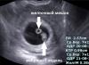
While waiting for the birth of a baby, a woman wonders at what time the embryo can be examined on an ultrasound scan? During the diagnosis for a period of 5-6 weeks, the fetal egg is about seven millimeters in diameter. At this time, in most cases, the doctor already visualizes the embryo. Around this time, you also manage to hear how his heart beats.
If you have a regular menstrual cycle, an embryo should be visible at the end of the sixth week. If the ultrasound does not show the embryo, it is recommended to undergo a second examination in a week to exclude all possible deviations.
There are also cases when the fertilized egg is outside the uterus. During ultrasound, the egg is not visible well enough, or it is not visible at all. In this case, the heartbeat is heard outside the walls of the uterus.
What to do if the fetus is not visible on the ultrasound and what can this mean?
There are situations that during the ultrasound scan, the embryo is not visualized inside the fetal egg, and sometimes the fetal egg itself. First of all, try not to panic. Maybe there is no pregnancy at all, or there was an error with the calculation of its term, so it is still difficult to diagnose. If the frozen pregnancy is not exactly confirmed, there is no need to rush to clean. First, it is better to undergo an ultrasound scan again, in another clinic. It may be necessary to conduct one or more studies. The best option is when, in parallel with the diagnosis, the level of hCG in the blood is monitored. If the pregnancy develops without deviations, then its level increases. This helps specialists to exclude a possible frozen pregnancy.
If an ultrasound does not show an embryo in a fetal egg, what does this mean?
Very often, a fetal egg without an embryo is diagnosed in the uterine cavity in young and healthy girls. Why is the fetus not visible on ultrasound, and is it possible to avoid a missed pregnancy?
There are many reasons for this phenomenon. This can be caused by infections of various etiologies, exposure to toxic substances, etc. on the body. You can minimize the possibility that an embryo will not be visible on an ultrasound by planning your pregnancy in advance in order to accurately calculate the gestational age. Also, you need to undergo examinations and, before planning the conception of a baby, cure all existing infections. This is especially important for women who are planning a pregnancy over the age of 35. This category has a significantly higher risk of chromosomal abnormalities in the fetus.

The absence of an embryo in a fetal egg often does not give a woman any signs during pregnancy. There may be bleeding if a miscarriage has begun. Even a gynecologist during the examination will not be able to say for sure whether there is an embryo in the fetal egg, or whether it is empty. The diagnosis of anembryony can only be made by a doctor who performed an ultrasound examination for a period not earlier than 5-6 weeks. If the gestational age is considered from the first day of the beginning of the last menstruation, then the doctor can visualize the embryo on ultrasound at 1-2 weeks of delay.
It is extremely rare for a patient to be misdiagnosed after an ultrasound, therefore, if there is no embryo in the fetal egg, it is necessary to check the result in a week on other equipment if there are doubts about the professionalism of the doctor or the quality of the ultrasound machine. An error is also possible for other reasons: a short gestational age or late ovulation, a woman's overweight and.
Why can't you see an embryo on an ultrasound?
If the pregnancy test shows two stripes, and the embryo is not visualized on the ultrasound, the reason for this may be:
- Incorrect calculation of the gestational age from the moment of conception. The embryo may not be visible because the woman is doing the test too early.
- Ultrasound diagnostics was carried out on an old apparatus or the specialist did not have the proper level of qualification.
- The study was done through the abdomen, not transvaginally.
- The pregnant woman had a miscarriage, but she did not pay attention to it (confusing it with the onset of menstruation), while the level of hCG in the blood had not yet decreased to its previous value.
If the ultrasound does not show the embryo in the fetal egg, do not immediately panic. For a number of reasons, the diagnosis of anembryonia can be erroneously made, so it is necessary to control the level of hCG in the blood and undergo the diagnosis again.
When a desired pregnancy occurs, all expectant mothers want to reliably make sure that the fetal egg is attached to the wall of the uterus and the formation of the unborn baby is normal. Ultrasound is considered the most reliable and convenient way to confirm a positive pregnancy test.
Despite the fact that a high-precision test strip, readily available in the pharmacy network, shows the onset of pregnancy, and a qualified obstetrician-gynecologist is able to recognize the symptoms of a "pregnant uterus", only the final ultrasound data confirms the fact of gestation. That is why, in the case when a woman believes that she managed to get pregnant, and the fetal egg is not visible on the ultrasound, future parents are perplexed.
In connection with this phenomenon, they have a question - can the diagnostician not see the pregnancy on an ultrasound scan? In our article, we want to provide information on how long it is possible to confirm the completion of the conception process, when the ultrasound scanner will allow the doctor to see the embryo, and whether it is possible not to see the pregnancy on ultrasound.
How are expectant mothers examined?
If the pregnancy test turned out to be positive, this can be confirmed by ultrasound - the diagnosis is carried out in a commercial center or in a antenatal clinic. It is important to know that an important role in obtaining reliable survey results is played by equipment with a high level of resolution and functionality, as well as the qualifications of a specialist.
Up to 9 obstetric weeks, two methods are used to examine pregnant women:
- Transabdominal - through the region of the anterior abdominal wall.
- Transvaginal - using a transducer that is inserted into the vagina.
Up to 5 weeks, the formed fetal egg is very small - its size is only about two millimeters. It is transvaginal that is considered to be an effective method for diagnosing the embryonic period - its high-frequency sensor makes it possible to get as close as possible to the uterine cavity and transfer the smallest sizes of the organs under study to the monitor screen.
The technique of examining a future mother using high-frequency waves is non-invasive and absolutely harmless - it allows the doctor to safely observe the development of the fetus
For the entire period of gestation, a woman performs at least three ultrasound scans. The examination session is short-term, the doctor tries not to hold the sensor in one place for a long time, especially during the formation of the most important organs and systems of the unborn baby.
What is seen on an ultrasound?
The main purpose of ultrasound in the embryonic period is to confirm the onset of pregnancy, this issue is especially relevant in the case of in vitro fertilization. The doctor-diagnostician has several tasks:
- Confirmation of fixation of the fetal egg in the uterus.
- Exclusion of the presence of a neoplasm in the uterine cavity, which can "mask" as pregnancy.
- Embryo viability assessment.
- Exclusion of ectopic pregnancy.
- Determination of the presence of a second fetus.
- Study of the localization of the placenta and fetus.
- Specification of gestational age.
In gynecological practice, there is one important point that all future mothers should know: the doctor measures the duration of the pregnancy period in obstetric weeks - from the first day of the last menstruation. That is why the difference between the real and obstetric term for conceiving a child is two weeks. In a woman of reproductive age with a normal menstrual cycle, recognition of pregnancy during transvaginal examination occurs no later than five weeks. If the cycle is irregular, it is difficult to determine the exact period for menstruation.
At what time is the embryo not visible on ultrasound?
Signs of a viable pregnancy are the following factors that the ultrasound scanner captures:
- the presence of a distinguishable outline of the embryo in the egg;
- listening to the fetal heartbeat;
- fixation of the slightest movements of the embryo.
For each woman, the period of bearing a child proceeds individually and it is very difficult to say exactly how long it takes for the doctor to be able to examine the fetus in the form of a dot and hear the rhythm of his heart.
In obstetric practice, there are certain normative terms for conducting ultrasound diagnostics for pregnant women. This takes into account that transvaginal scanning allows you to study the ongoing changes before transabdominal. In order for our readers to evaluate the quality of these methods, we provide a comparative table.
The beginning of contractions of the heart muscle of the future baby falls on the period from 3 to 4 weeks and it is possible to catch it only with the help of a transducer (a special narrow vaginal sensor). It happens that the uzist doctor cannot see anything in the fetal egg and recommends coming for an examination in 7-14 days.
It is the frequency of contractions of the heart muscle of the embryo that will allow the doctor to clarify the gestational age:
- at 5 obstetric weeks, the heart rate is up to 85 beats / min;
- in 6 - from 102 to 126;
- in 7 - from 127 to 149;
- in 8 - from 150 to 172;
- at 9 - 175.
If at 7 obstetric weeks no embryo parameters are observed in the fetal egg and the heart rhythm is not heard, a preliminary diagnosis of anembryony is made - the absence of an embryo in the fetal egg. However, in this case, the woman is also recommended to come for an additional ultrasound after another 7 days.
Embryo parameters
Normally, the fetal egg has an oval shape and a dark gray tint. To fully monitor the formation of the fetus on ultrasound, the following indicators are measured.
Many factors influence the clear visibility of the fetus on the monitor of the ultrasound machine, and if the embryo is not visible, do not panic - you should wait two weeks and repeat the study.

At the beginning of pregnancy, the embryo resembles the letter “C”, as it grows, the appearance changes - at 8 weeks you can already see both the head and the highlighted limbs
Why is the fetus not visible on ultrasound with a growing level of hCG?
The fetal membranes of the developing baby produce a special substance - human chorionic gonadotropin, indicating that the conception has taken place. In the first trimester, the amount of this protein-hormone in the circulating blood of a woman grows very quickly - in the first weeks, its concentration doubles every second day.
Monitoring the growth dynamics of hCG levels allows obstetrician-gynecologists to draw an accurate conclusion about the development of pregnancy.
If, when assessing the amount of this biologically active substance, an increase in its amount is observed, the doctor confirms with certainty the onset and successful development of pregnancy. Every woman wants to know about the onset of pregnancy early, but the accuracy of the ultrasound results in the second week of the delay in menstruation is very low - it is better to wait until the fifth week.
If, with positive hCG tests (in the case when the quantitative final data of the analyzes correspond to the estimated gestational age), pregnancy is not determined by ultrasound, then you need to come for an additional examination. An hCG level of more than 1800 mU / ml corresponds to the third week of pregnancy and, if an ultrasound scanner does not observe a fetal egg in the uterine cavity, the doctor assumes the development of an ectopic pregnancy.
The lack of growth in hCG levels (negative test) may indicate the fact that the development of the embryo does not occur - either it died, or the egg was not fertilized in this cycle.
Not all women know such a phenomenon as biochemical pregnancy or preclinical spontaneous miscarriage. In this case, conception occurs, the fetal egg is attached to the uterine wall, however, when the next period comes, the pregnancy is terminated.
Emphasis should also be placed on those situations where pregnancy is not visible on ultrasound, and the test is positive - monitoring the level of hCG is of particular importance, it is necessary to take a blood test several times, with an interval of several days. The final data of laboratory studies allow us to determine the compliance of the hormone concentration with the norm and its increase.

Practitioners advise future parents to try not to force events, an exception is possible only when it is necessary to confirm or deny the pregnancy as soon as possible
What to do if pregnancy is not detected during ultrasound scanning?
In the event of a situation where the uzist doctor cannot see the outlines of the embryo, and sometimes the fetal egg itself, you must try to remain calm and not succumb to false beliefs! This is possible in the absence of gestation or its period is very short to notice on the monitor. Without the presence of absolute evidence of an interrupted pregnancy, it is impossible to carry out curettage of the uterine cavity!
You should go to another clinic and re-examine - it is better to do this on expert-class equipment with high resolution. It is also necessary that the ultrasound is accompanied by blood tests for hCG levels. You may need to go through the examination several times. Future parents should make every effort so that diagnostic errors do not cost the child's life!
2015-12-25 16:27:48
Catherine asks:
Background: On November 21, there was a burning sensation and blisters in an intimate place. On November 26, I passed the PCR test for Herpes type 1 and 2, it came positive (on November 26, I immediately started treatment with acyclovir. But on December 11, there was a delay, I took a test ... I was pregnant! .e at the time of infection if even the primary embryo was only 5 days old.
On December 16, I donated blood by ELISA for Herpes 1 + 2 LgM-positive ((I’m just in a panic, because I don’t remember if I ever had genital herpes at all, I didn’t pass on LgG.
do not panic and wait for the results on G? And how then to understand whether it was primary or relapse? If relapse then M + G- should be? And if the primary infection, could the child already when it had not yet formed to become infected with this? I did an ultrasound on December 15, the embryo was not visible, just a sac was set for a period of 3-4 weeks. On December 24, I did a second ultrasound, the embryo is already visible and the heart is audible, the period of 6 weeks is set. And how long should the positive M remain?
Responsible Hovhannisyan Karine Eduardovna:
Hello Ekaterina! First, I want to reassure you. The fact is that the herpes virus is detected in more than 80% of women, and if all of them developed a pathology in the fetus, then the human race would no longer exist. But still, you need to make sure. There are antiviral drugs that increase immunity and that can be taken by a pregnant woman. Such drugs are prescribed by the doctor with whom you will be registered for pregnancy. These drugs are safe for both the pregnant woman and the unborn child. In addition, it is necessary to do a screening of the pregnant woman in due time, which will completely dispel your fears. The only thing is to carry out antiviral treatment in order to prevent exacerbation during childbirth, otherwise, you will have to do a caesarean section so that the child does not become infected during passage through the birth canal.
2012-11-11 16:07:21
Alina asks:
Please tell me, I have 7 weeks of obstetric period, 4-5 weeks of embryonic. the ultrasound does not show the embryo, the fetal egg is 19 mm ... what does it threaten?
2008-12-20 12:12:35
Oksana asks:
Hello, tell me, please... According to my calculations, the term from conception is 3.5-4 weeks... The ultrasound does not show the embryo... the doctor says almost with certainty that there is no embryo. Pregnancy does not develop ... Is this possible at such a time?
Responsible Filippova Olga Yurievna:
The gestational age is considered: the moment of conception + 7 days for implantation (ingress) of the fetal egg into the uterine cavity. Uterine pregnancy on ultrasound by the transabdominal method (through the abdomen) can be visualized in a period of 4-5 weeks. So count.
2016-05-04 14:37:28
Eugene asks:
Hello!
I have the beginning of the 7th week from the day of the last menstruation or the 5th week from the day of ovulation (day 17 of the cycle) The fetus is not visible on the ultrasound (it was checked on 4 devices). The doctor advises to do a purge, as it suggests a threat to my health (but only because the embryo and pregnancy hormones 22000 are not visible). Blood tests are in order, I take folic acid, calcium and hormones. Yesterday, according to the doctor, I should have bled and I went to the ambulance, but everything turned out to be in order. No bleeding. I decided to wait a week and a half. If at least one chance that the pregnancy will not terminate?
Responsible Bosyak Yulia Vasilievna:
Hello Evgeniya! First of all, I advise you to take an analysis for hCG in dynamics, every 2 days. With a normally developing pregnancy, the indicator should double. With an ectopic pregnancy, the progression of hCG growth will be underestimated; when fading, the indicator does not increase or falls. Honestly, you should not hope for the normal development of uterine pregnancy. First of all, it is necessary to exclude an ectopic pregnancy.
2015-12-24 17:38:09
Catherine asks:
Background: On November 21, there was a burning sensation and blisters in an intimate place. On November 26, I passed the PCR test for Herpes type 1 and 2, it came positive (on November 26, I immediately started treatment with acyclovir. But on December 11, there was a delay, I took a test ... I was pregnant! It turns out that the last menstruation was on November 12. Estimated conception is 16.11 or 19.11.
On December 16, I donated blood by ELISA for Herpes 1 + 2 LgM-positive ((I’m just in a panic, because I don’t remember if I ever had genital herpes at all, I didn’t pass on LgG. I’ll run tomorrow morning. I read everything to tears ( (
Girls tell me who did it?! do not panic and wait for the results on G? And if the primary infection, could the child already when it had not yet formed to become infected with this? I did an ultrasound on November 15, the embryo was not visible, just a sac was set for a period of 3-4 weeks. Today I did a second ultrasound on December 24, the embryo is already visible and the heart is audible, the period of 6 weeks is set.
Responsible Wild Nadezhda Ivanovna:
Herpes infection occurs with periods of exacerbation and remission. In this case, infection and an acute process of genital herpes were possible. It is bad that there was an acyclovir intake - for the embryo, but treatment is needed for the herpes infection itself in a pregnant woman. You will be advised to terminate the pregnancy, after which you need a good course of treatment together with your sexual partner. In case of pregnancy, you must realize that you need prenatal screening of the 1st and 2nd trimester of pregnancy, a consultation with a geneticist. It is desirable to take proteflazid (phytopreparation with antiviral effect) or engistol, periodically - viferon suppositories. A man should undergo a good course of anti-inflammatory therapy, sex only with a condom, or abstinence. Ultrasound at 19-21 weeks on a 3D machine. During pregnancy, control over the course of herpes infection: lgM, lgG, PCR smear for herpes type 2, cytological smear, colposcopy at least 2 times. During pregnancy, infection of the fetus is possible - there is always a risk, most of all in the third trimester and in childbirth, this is with the condition that in the first trimester it is lucky and the fetus develops. The choice is yours, if pregnancy continues to develop - be optimistic, doctors do not scare - they talk about the worst and warn. But, the choice is always up to the couple or the woman herself. Good luck.
2015-07-31 18:08:32
Tansholpan asks:
Hello, my long-awaited pregnancy, but something went wrong, on May 20 I had my last menstruation, ovulation was late from about June 16-21 for 4-5 weeks, I went for ultrasound, they wrote a fetal egg in the uterine cavity, cf 23.9 ktr 7.3 and a yolk sac size did not write the embryo no. after a week I went to another ultrasound 5 6 weeks the embryo is missing, she said that the fetal egg is growing and the yolk sac needs to have an abortion because you are more harmed, my question is whether the embryo is still growing? what am I supposed to do?
2013-07-03 18:07:59
Julia asks:
Hooray, it worked!!! I wrote to you earlier that I could not get pregnant for 8 years. I donated blood for hcg, it turned out 4840 units. But on ultrasound after 3 days they did not see the embryo, on the left ovary there is a cyst of the corpus luteum and the ovum is deformed, the pregnancy is uterine. I was reassured and appointed to redo the ultrasound in another 4 days in order to exclude anembryony. The last menstruation was 18.05. and 19.06. just a little smeared. It seems to me that I am at the end of the cycle and the period is really short. Now I have a cold, a runny nose and a sore throat, the temperature is 36.4-37.4. I was prescribed arbidol and drops in the nose, brown discharge began a little. Is it dangerous for the baby and at what time should the embryo be visible? Thank you.
Responsible Gritsko Marta Igorevna:
The embryo should be clearly visible from 5-6 weeks of gestation, heartbeat from 7 weeks. I advise you to treat a cold with folk methods - frequent gargling, warm, plentiful drink (linden, raspberry, etc.), Pinosol can be used from drops. Over the next week, I think the situation with the course of pregnancy will be completely cleared up. I wish you success!
2012-10-10 10:41:18
Inna asks:
Good afternoon! I have a question for you, my last menstruation was on 25.08.2012, 02.10 I went to the gynecologist with a square egg of 3 mm, they said to come in a week for a follow-up ultrasound. I came on 09.10, they did an ultrasound, they said that the square of the egg had grown to 7.5 mm, but the embryo was not visible, they said to come in another week, please explain to me if this is some kind of pathology or a short time, even I can’t understand anything.
Ultrasound is an important diagnostic procedure for gynecological examination. It is especially important for monitoring the development of the fetus. Thanks to ultrasound diagnostics, it became possible to visually assess the embryonic development of the child, determine the compliance of the development of the baby with the estimated gestational age. The doctor can determine not only the level of development of the fetus, its size, but also find out what condition it is in, i.e. identify his possible suffering (for example, hypoxia). This helps obstetricians-gynecologists to form the right strategy for managing pregnancy. Ultrasound, as a diagnostic method, allows you to confirm the very fact of pregnancy.
Ultrasound diagnostics can confirm or deny the fact of pregnancy with almost one hundred percent probability.When can pregnancy be confirmed by ultrasound?
It makes no sense to run for an ultrasound the next day after unprotected sex. The size of the egg, even a fertilized one, does not allow it to be seen with ultrasound imaging. How long does it take to see the fetal egg and determine its size? You can see the embryo when the fetal egg is at least 1 cm. If there was a delay in menstrual bleeding for a week, by this time the gestation process has approximately reached 6 weeks. By this period, when diagnosing with a high-precision apparatus, the fetal egg is already visible. It is not yet possible to determine the presence of an embryonic heartbeat and consider its structure.
Due to the peculiarity of ultrasound diagnostics in the early period of gestation, namely the need to insert a transducer into the vagina, such an examination is carried out strictly according to indications. The grounds are the suspicion of ectopic fixation of the fetal egg, cystic drift.
There may be other considerations, based on which the gynecologist offers the woman this procedure.
When is a qualified, highly experienced specialist on a good device that provides high-quality visualization able to recognize the signs of pregnancy? After 3 weeks. The ectopic location and fixation of the fetal egg is diagnosed by ultrasound after 2 weeks (transvaginally) and on the 20th day after conception (through the peritoneum). To help the data of ultrasound diagnostics, a blood test is usually prescribed. HCG indicators make it possible to judge the implantation of the embryo. At 7-8 weeks (from about 10 days of delay), a good ultrasound diagnostician determines pregnancy with almost 100% certainty.
Why doesn't ultrasound "show" pregnancy?
If all signs of gestation are present, the level of gonadotropin indicates successful implantation of the embryo, and ultrasound data do not show a fetal egg and do not confirm the fact of pregnancy in a particular patient, this does not mean that the pregnancy did not take place. The reasons why an embryo is not visible on an ultrasound may be as follows:
- used transabdominal sensor;
- low accuracy of the equipment;
- incorrect calculation of gestational age, ultrasound diagnosis is carried out early, the fetal egg is not visualized;
- gynecological pathology (yellow cyst, for example).
For the expectant mother in this situation, the main thing is not to panic. It is necessary to repeat the hCG test (its indicators should double in 2 days - this is an indicator of a normally developing pregnancy) and re-visit the ultrasound room in a week.
 The use of a transvaginal probe is much more informative for detecting a normally located embryo than a transabdominal examination.
 What justifies the need for early ultrasound diagnostics
With a hCG result of 1000-2000 mU per liter, ultrasound can be effective. This study will help the obstetrician-gynecologist recognize possible early pregnancy problems and its rate. It could be:
- ectopic pregnancy;
- confirmation of the fact of embryo implantation;
- finding out the reasons for the delay in menstruation in the absence of pregnancy;
- establishing the gestational age (the smaller it is, the more accurate the data);
- determination of pregnancy fertility (not always possible);
- establishment of the threat of disruption.
Ultrasound diagnostics in the early period is carried out only in exceptional cases, but still it is better to wait 5-8 weeks. At this time, the embryo is visible and it is already possible to determine the rate of its development.
If the ultrasound was performed early, the suspicion of pregnancy is the size of the corpus luteum. With a delay in menstruation and a corpus luteum size of at least 16 mm, we can talk about pregnancy, although the fetal egg is not yet visible.
How long does ultrasound show and its accuracy?
The age of the fetus is determined by the obstetric method and embryonic. The first is counted from the 1st day of the last menstrual bleeding, the second counts the time of gestation from the day of conception (this date is considered the day of ovulation). The embryonic period is 2 weeks shorter than the obstetric one. In the ultrasound procedure, the obstetric reference method is considered the basis. The procedure itself is not a computational mechanism for calculating the gestational period. It consists in determining the degree of fetal development and correlating the data with the obstetric period. The accuracy of ultrasound (by determining the number of weeks of pregnancy) directly depends on the gestational age itself:
- up to 12 weeks - accuracy is 1-2 days;
- from 12 to 28 weeks - the error is a week in both directions;
- after 28 weeks, the error increases.
 After 12 weeks, the accuracy of determining the gestational age is significantly reduced.
 The term of ultrasound does not coincide with obstetric: reasons
A deviation of 14 days in both directions is not considered a pathology, obstetric practice allows this. For example, if the terms for ultrasound exceed the obstetric calculations in the initial period of gestation, the reason may be an error in determining the obstetric period, due to the fact that almost immediately after fertilization there was a small spotting, which the woman took for menstruation. The second reason may be the large size of the fetus.
When researching, it is necessary to take into account the heredity of the baby. Large parents may have large children, short miniature couples and children may be small. Also, the fetus on ultrasound may be smaller in size than it should be according to the estimated period, if the doctor recorded the embryonic period. This is where natural causes end.
The fetus may develop inappropriately with hypoxia or other pathologies. To clarify the condition of the child in the womb, the doctor prescribes dopplerometry. For a woman, this procedure is no different from the studies she has already undergone, but it allows you to clarify the diagnosis.
What to do if the size of the fetus does not correspond to the obstetric term?
Consult with your doctor. He will conduct an examination, determine the height of the uterus and measure the circumference of the abdomen, evaluate the indications of the mother's condition and suggest either hospitalization or re-examination in a week. There is no need to refuse hospitalization and re-examination, because not only the condition of the expectant mother, but also the life of her child depends on this. In the hospital, additional examinations can be carried out, which will either show that everything is fine, or help prescribe adequate treatment.
Ultrasound diagnostics is an art. Much depends on the qualifications of the doctor. A good specialist on precise equipment is able to determine pregnancy with a vaginal sensor for a period of 3 weeks or more (according to the obstetric method). But in fact, how many weeks the doctor can confirm the pregnancy depends on the individual case.