The genus Escherichia includes several species, but the undisputed leader among them is E. coli. This is the type species of the family Enterobacteriaceae, which combines gram-negative facultative anaerobic rod-shaped bacteria that do not form spores and have a number of other common features.
The generic name enshrined the priority of the German bacteriologist T. Escherich, who isolated E. coli in 1885 from the feces of a child with "infantile cholera", which was widespread in Europe. But the recognition of its morbidity was delayed for many years. The fact is that similar bacteria were invariably detected in the intestines of healthy children and adults, which contradicted one of Koch's postulates, which strictly distinguished between pathogenic and harmless microbes. The ubiquity of Escherichia coli is also recorded in the first name proposed by T. Escherich - Bacterium coli communis. Its role in the pathology of the intestine was not denied, but many were in doubt. More convincing was the participation of E. coli in extraintestinal pathology, especially in inflammatory diseases of the urinary system. A wide representation in the normal microflora served as a reason for recognizing the harmlessness of Escherichia coli. Attempts to change the situation, recognizing the pathogenetic heterogeneity of Escherichia, were perceived with interest, but did not lead to productive generalizations. A radical shift took place in the 1950s. after the Danish bacteriologist F. Kaufman substantiated the principles of serological classification (immunotyping) of Escherichia based on surface antigens.
Morphology.E.coli represented by straight gram-negative rods, 0.44-0.6x2.0-^-6.0 µm in size, motile due to peritrichial flagella. Some are characterized by the presence of a microcapsule built from a sialic acid homopolymer; such strains are referred to as TO + . "
cultural properties. On dense media, they form colonies in S- and R-forms. S-shaped colonies are smooth, shiny, translucent. On liquid media, they form diffuse turbidity and bottom sediment.
biochemical properties. It has a pronounced biochemical activity (see table. 16.9). The biochemical properties that form the basis of differential diagnosis during bacteriological examination are as follows:
production of acid and gas during the fermentation of glucose,
lactose fermentation,
inability to form hydrogen sulfide,
indole production.
Antigenic structure.E.coli has a complex antigenic structure:
a) has a somatic O-antigen that determines the serogroup. About 171 varieties of the O antigen are known;
b) surface K-antigen can be represented by 3 antigens: A, B and L, differing in sensitivity to temperature and chemicals. In Escherichia, more than 97 varieties of the K-antigen are found, mainly of the B type. The K-antigen has the ability to mask the O-antigen, causing the phenomenon of O-inagglutinability. In this case, the O-antigen can be detected only after the destruction of the K-antigen by boiling;
c) the type-specific antigen is the H-antigen that determines the serovar, of which there are more than 57.
The antigenic structure is indicated by the formulas of the serogroup as O:K, the serovar - O:K:N, for example: 012:B6:H2.
Usually, only O-group affiliation is identified, since it is this feature that best agrees with pathogenicity (although it does not explain it!).
Strain diversity in combination with host reactivity determines the eco-pathogenetic strategy of Escherichia coli:
(1) non-pathogenic (residenttnye) commensals that colonize the large intestine for life;
(2) Escherichia, callinging extraintestinal lesions - from cystitis tosepsis; These infections usually occurendogenously, due to strains that colonizeintestines;
(3) diarrheagenic Escherichia, whichare of exogenous origin, do not delayvayutsya in the body (transient strains) and,from this point of view, can be considered a pathogennye varieties of Escherichia coli.
resistance. It remains in water and soil for several months. Dies when heated to 55 °C for 60 minutes, at 60 °C - for 15 minutes. Escherichia in the environment are able to go into an uncultivated form
Ecology, features of distribution and pathogenesis. View E.colint is homogeneous, but is divided into subspecies. There are opportunistic Escherichia and diarrheagenic.
Opportunistic Escherichia are part of the intestinal and vaginal microflora in humans. E.coli also make up the intestinal microflora of mammals, birds, reptiles, and fish. With feces, the microbe is released into the environment. The presence of E. coli in water, soil, food, household items is an indicator of fecal contamination.
Conditionally pathogenicE. coli are you capablecall endogenous purulent-inflammatoryprocesses of various localization, calledgiven by parenteral escherichiosis.Parenteral escherichiosis can besepsis, suppuration of wounds, secondarypneumonia, urinary tract infectionsways. Often occurs against the background of immunodevelopmentficitta,
Strains E.coli, involved in the infectious process of the lower urinary tract, have a specific O-antigen that allows them to adhere to the surface of the bladder epithelium. Those strains E.coli, which cause an infection of the upper urinary tract (pyelonephritis), have special P-fimbria with antigenic properties. These P-fimbria are
allow the microbe to adhere to the epithelium of the collecting ducts. E.coli, causing urinary tract infection, more often belong to serogroups 02, Gb, 09. Some of them have hemolytic activity due to the presence of the H1y-plasmid.
The vast majority (about 80%) of neonatal meningitis are caused by E.coli, which the newborn becomes infected through the birth canal. E.coli, causing neonatal meningitis often has a microcapsule composed of a sialic acid homopolymer. The presence of a microcapsule gives the pathogen antiphagocytic properties, since the microbe ceases to opsonize due to the loss of the ability to activate complement.
Of the opportunistic E.coli strains multiresistant to antibiotics can be formed due to the acquisition of R-plasmids, which become causative agents of nosocomial infections.
Pathogenic E.coli, which are the causative agents of intestinal escherichiosis, AII, are called diarrheagenic.
ACUTE INTESTINAL INFECTIONS
Infection with Escherichia coli occurs immediately after birth and ends with asymptomatic colonization of the large intestine, where Escherichia dominate among the aerobic (more precisely, facultative anaerobic) microflora. Infection with most strains in later life also goes unnoticed. Much less often, acute (diarrheal) intestinal pathology develops - escherichiosis or intestinal coli infection. diarrheagenicity(i.e., the ability to infect the intestinal epithelium) was recorded in representatives of many O-groups, although not all strains within each of them have this ability. O-antigen marks the potential pathogenicity, the implementation of which requires additional factors.
epidemiologist and I.
Escherichiosis is a disease anthroponotic withfecal-oral transmission mechanism. Exception constitute enterogem orrag logically e Escherichia, the reservoir of which is, apparently, cattle. Other diarrheagenic Escherechia also circulate among animals, birds, and even insects (cockroaches), but this is unlikely to be of epidemic importance.
The leading role belongs
Dietary transmission, primarily through milk and dairy products.
The second most important is the waterway.
Contact-household transmission (household or child care items, hands of mothers, staff of child care facilities and hospital hospitals) is possible when infants are infected with EPK strains (see below).
■ The aggressiveness of diarrheic Escherichia is not high. P atogenetically significant dosage is 10 6 -10 9 bacteria, which is several orders of magnitude higher than that of salmonella typhoid and shigella. Susceptibility is highly dependent on strain characteristics and is much higher in young children, especially the first year of life. ._ Acquired age_agestnoy resistance may be associated with vaccine(protective) effect of Escherichia-comensals that have common antigens with virulent strains, as well as with subclinicalby infection with diarrheagenic Escherichia-mi.
Incubation period usually 1-3 days and never more than 7 days.
chief source of infection patients serve (often with erased forms of the disease), excreting a huge amount of the pathogen with feces. Of less importance are convalescent cents and bacteria carriers. This is due to the fact that carriage rarely lasts more than 2-3 weeks, and the amount of bacteria isolated is so small that it does not pose a direct threat. Dangerous is the contamination of food, in which E. coli finds suitable conditions for reproduction. This explains the endemic spread of escherichioses in hot regions with a low level of sanitary culture. In industrialized temperate countries, coli infection is rare.
Based on the pathogenetic mechanismsmov, serological markers and epidemiological features differentiate fivevarieties of diarrheagenic Escherichia:
(1) enterotoxigenic,
(2) enteropathogenic,
(3) enteroinvasive,
(4) enterohemorrhagic and
(5) enteroaggregating (enteroadhesive).
Enterotoxigenic Escherichia coli(ETKP) were found among representatives of more than 70 O-groups, more often among O-groups, 078, 0128 and 0153 (in total they make up more than half of ETCH). ETECs produce toxins that disrupt the balance between secretion and absorption of fluid by the epithelial cells of the small intestine. Excess water and electrolytes in the intestinal lumen causes diarrhea. Various forms of the disease are possible - from mild diarrhea to cholera-like intoxication ("minor cholera"). Children and adults get sick. Only in rare cases (more often in premature newborns) does dehydration require emergency care (rehydration therapy).
The spread of bacteria is limited to the surface of the mucous membrane, enterocytes are not invaded and are not structurally damaged. This is evidenced, in particular, missingeffect of the inflammatory response in the intestinal wallnika and watery stools without admixture of slizee and blood. This type of diarrhea is called "secretory", in contrast to the invasive variant, which is observed during the destruction of the intestinal epithelium. Secretory diarrhea occurs due to the hypersecretion of Na\CI and water by the crypt border cells with a weakening of the alternative function of the small intestine - the absorption of electrolytes and water by the villous cells of the mucosa.
The most well-known are the two escherichial enterotoxins - thermolabile (LT) And thermostable (ST). They are encoded in plasmids - discrete or general. In the latter case, both toxins are synthesized simultaneously (about 5% of strains).
LT form approximately 25% of ETEC strains. According to the mechanism of action (ADP-ribosyl transferase) A-B toxin and even in antigenic properties, it is similar to cholerogen - the enterotoxin of cholera vibrio. After reception by gangliosides A Enterocytes LT (more precisely, its A-subunit) penetrates the cell and inactivates (ADP-ribosylates) the regulatory protein that controls the activity of adenylate cyclase. The diarrheagenic effect is associated with an increase in the intracellular level of cyclic adenosine monophosphate (Fig. 2).
ST (produced by about 70% of ETEC strains) looks and acts differently, although from a pathogenetic point of view it does a “common cause”. It consists of two low molecular weight peptides (StA and StB) that are weakly immunogenic and resistant to heat, proteolytic enzymes and acids. ST does not enter the cell by acting on enterocyte receptors associated with membrane guanylate cyclase. Strengthening the synthesis of cyclic guanosine monophosphate disrupts intracellular. balance in cyclic nucleotides, provoking hypersecretion of water and electrolytes.
Toxin formation is preceded by fixingETEC on enterocytes especially in the proximal small intestine. This requires non-standard paraphernalia, which are deprived of the "ordinary" E. coli inhabiting the large intestine. Among the factors that provide selective colonization of enterocytes, the best studied are CFA/ I, CFA/ IIAndCF/ IV(from English.colonizationfactorantigens). They are approximately equally distributed between the ETCP strains and represent lectins 1 Lectins are proteins that selectively bindcalling certain carbohydrates (from lat.legere- knock outarmy), structurally formed in the form of pili/fimbriae (Fig. 3). According to one of the general classifications, it is accepted differentiate mannose sensestel and mannose-resistant adhesins escherich. The former interact with the mannose radicals of glycoproteins and are therefore blocked by mannose and its derivatives, the latter do not react with mannose. CFA are mannose-resistant, which distinguishes them from the vast arsenal of fimbriae and non-fimbriae factors involved in "banal" colonization.
large intestine. In contrast, CFAs are taken up by the same cellular gangliosides (GM1) that fix LT. Note that fimbriae of animal ETCs (K88, K89, 987P) are also resistant to mannose, although they are not identical to the adhesins of “human” ETCs. This explains the reason for the anthroponism of enterotoxigenic co-infections.: ETC of animals are not able to colonize the human small intestine, which, by the way, is also true for other categories of diarrheagenic Escherichia (see below). Enterotoxins of human and animal ETECs are also structurally non-identical, although they are similar in their mechanism of action.
CFA encoded in plasmids "by neighbor to the genes of enterotoxins, and only the simultaneous expression cfa - And tox -genes provides ETCP-virulence . Without colonizing factors, enterotoxins are pathogenetically inert, just like CFA-adhesins without toxigenicity. The study of factors of colonization is ongoing. This is prompted by observations of ETEC strains lacking CFA: they account for about 20% of isolated cultures. By the way, C F / II and CF / IV are not homogeneous in themselves. Each of them consists of three antigens - CS1, CS2, CS3 (CRAL1) and CS4, CS5, CS6 (CFA / IV) (from the English cell surface). SRL is structurally homogeneous.
ETEC remains the leading cause of sporadic and epidemic childhood diarrhea in tropical and subtropical developing countries. In developed countries, ETES rarely manifest itself, mainly in the form of "traveler's diarrhea", which is observed in tourists visiting regions that are unfavorable for ETES infection.
Enteropathogenic Escherichia coli(EPKP). From them in the 1950s. the modern stage in the study of diarrheagenic Escherichia started. This explains the unfortunate universalism of the term (enteropathogenicity), which can equally be extended to all diarrheal-gene Escherichia. EPCs, which include representatives of about 20 O-serogroups (usually 055, 0111,0119, 0127, 0128), cause damage to the small intestine in young children (up to two years, usually in the first year of life). Previously, the disease was called "toxic dyspepsia."
EPCs affect the small intestine. The diarrheal syndrome is based on the rearrangement of intracellular homeostasis of enterocytes, which is excited by signals from cell receptors occupied by EPK adhesins. The result is smoothing of microvillifor the accumulation of filamentous actin in the zonecytoplasm adjacent to the site of the primarybacteria adhesion. This leads to impaired fluid absorption, initiating secretory-type diarrhoea (see above). Among the main adhesins are the outer membrane proteins encoded in the chromosome, called « intimins" 1 "The term emphasizes exceptionally dense("intimate") contacts of bacteria with cells, devoid ofmicrovilli, and derivatives of plasmids with a molecular weight of 60 mD - a mandatory attribute of EPKP.
In general, in terms of their pathogenetic consequences m EPCP-adge zia, unique, having received the name mechanism"attach-smooth I " (eng.attaching-effacing).
EPCO-diarrhea are classified as enteritis, although pathogenetically this is not true. Enterocyte damage is functional and, like enterotoxigenic diarrhea; Not resistance They multiply in the epithelial cells of the colon and, damaging them, induce inflammation and ulceration of the mucous membrane. Enteroinvasiveness is limited to representatives of several O-groups: 028,0112,0124,0136,0143, 0144, O152, O164. They are responsible for a small percentage of all cases of bacillary dysentery, significantly inferior to shigella. But this is enough to make the problem tangible in absolute terms, especially with a low level of sanitary culture. EIEC usually affects children either sporadically or in outbreaks in organized communities. Adults rarely suffer - during water or food epidemics.
EICP, Enteroinvasiveness reflects similarities between Escherichia and Shigella. The homology of their DNA exceeds 90%, which is quite enough to unite at the level of the genus and even the species. Such attempts have been made repeatedly, but practical considerations and the strength of tradition have prevailed over academic claims, ensuring the permanence of these two classical groups of enterobacteria. At the same time, with such a close relationship, intermediate forms should be expected that share the properties of Escherichia and Shigella. Most of the enteroinvasive strains of Escherichia coli belong to such “hybrids”. Many of them were discovered due to their "atypicality", i.e. the presence of signs common with shigella: immobility, anaerogenicity (fermentation of carbohydrates without gas), slow fermentation of lactose or the complete absence of this sign, relatedness by O-antigens. Their invasiveness is judged by the ability to cause keratoconjunctivitis when a bacterial suspension is instilled into the conjunctival sac of a guinea pig or by a cytopathic effect in epiphyte cultures. telial cells. Taxonomic doubts are eliminated by expanding the set of biochemical tests. In a number of ways (production of lysine decarboxylase, fermentation of citrate, utilization of sodium acetate as the only source of carbon), such cultures differ from Shigella and are considered as atypical Escherichia. Classical (i.e. fast paced zl ag ayu ing lactose) strains are sometimes also enteroinvasive, although careful analysis can reveal signs of phenotypic similarity to shigella (for example, the absence of lysine decarboxylase).
The similarity to Shigella extends to the mechanisms that ensure invasiveness. They are controlled by plasmid genes homologous to Shigella virulence plasmids. The effectors of invasion are not exactly known, but their analogy with Shigella factors is undoubted. Invasion begins with the penetration of M-cells of the intestinal epithelium and evolves through lateral contacts of epithelial cells. Inside the cells, bacteria produce enzymes that lyse the phagosome wall, causing damage to epitheliocytes and the spread of infection.
Enterohemorrhagic intestinal coliki (EGCP).From the point in terms of virulence, this is perhaps the most equipped variety of Escherichia. Most EHECs belong to the O157 serogroup (O157:H7 serotype), less often to 026, Olll, O145. EHECs colonize the large intestine (especially the cecum), causing hemorrhagic colitis . Unlike sh igellosisnyh and e ANDKP-lesions bloody is Prague-neni I wouldsingle leukocytes. W The disease is accompanied by general intoxication (nausea, vomiting), and in the most severe (fortunately, rare) cases, extraintestinal symptoms, the most formidable of which is hemolytic-uremic syndrome (hemolytic anemia,thrombocytopenia, acute chronic insufficiency). However, often the infection is mild, possibly a healthy carriage.
The initiating role belongs to the aggressive intimindependent adhesion, commented above on the EPKP model. It is followed by production of toxins which have a local effect (hemorrhagic colitis) and cause systemic effects (damage to the kidneys, central nervous system, intravascular hemolysis). Several cytotoxic EHEC factors are known, which are easy to detect by hemolysis, although this is only one of the manifestations of their membranotropic (cytolytic) activity. . Excepthemolysins , produced by strains of many O-groups, EHECs secrete specific iecytolysins. They are called verotoxins”(according to the toxic effect in the Vero cell line culture”) or “shigo-like toxins”, implying similarity (primarily neurotropism) with the Shigella dysenteriae toxin. Special, enterohemorrhagic hemolysins are also described. Perhaps we are talking about the same factors, although the complex * toxin spectrum of EHEC is not in doubt. One can only think about the pathogenic leadership of individual toxins, not forgetting the likelihood of a cooperative effect. EGCP-cytolysins are of a plasmid nature. Plasmid genes are also required for intestinal colonization. Like EPKD, enterohemorrhagic strains carry a plasmid (p!5 m.m. 60 mD), the products of which, along with inlmin and another 94 kD outer membrane protein, are involved in pathogenetically important adhesion.
There is reason to believe that, unlike other co-infections, EHEC-escherichioses are zoonoses. The most likely source of infection is cattle. This is evidenced by a number of outbreaks of O157-escherichiosis recorded in Canada, the USA and Japan. Infection occurs by eating meat (after insufficient heat treatment), as well as raw milk. However, not everyone is in a hurry to abandon * anthropogenicity, preferring to talk about the anthropozoonotic nature of the infection. Another paradox: children of the first year of life do not get sick with EHEC-escherichiosis. Perhaps this is also the result of the epidemiology of this infection, which is unusual for escherichiosis: newborns are maximally protected from contact with "zoonotic microbes".
Enteroaggregating (enteroadhesive) Escherichia coli (EACP). An additional reason for complicating the classification of diarrheal Escherichia was the morphological features of their adhesion in epithelial cultures! human cells (HEp-2 and HeLa). One of the variants of adhesion, resembling brickwork (stacked-brick), is due to the fixation of bacterial aggregates on the cell membrane (Fig. 5). Such strains are allocated to an independent category of diarrheagenic Escherichia and are considered one of the causative agents of diarrheal; (especially stubborn) syndrome in children. EACP colonize various parts of the intestine and produce several cytotoxins. The structural basis and pathogenetic significance of EACP adhesiveness are unknown. It is also not clear why, having a wide ability to colonize the intestinal epithelium, EACP affect mainly the large intestine.
ETCP are causative agents of cholera-like diseases in children and adults.
Pathogenicity is determined by the production of heat-labile (LT), structurally and functionally related to cholera toxin, and heat-stable (ST) enterotoxins determined by the Ent-plasmid, and colonization factors CF (colonization factor, English), the synthesis of which is also determined by plasmids. Thanks to CF, ETEC proliferate on the surface of the epithelium of the small intestine (see Table 16.10). ETEC colonization of the surface of the small intestinal mucosa provides a massive release of enterotoxins that disrupt the water-salt metabolism in the intestine, leading to the development of watery diarrhea. The mechanism of development of diarrheal syndrome is associated with the activation of LT intestinal adenylate cyclase, and ST - guanylate cyclase. 17 serogroups are associated with ETEC, among them the serovars O6:H16, O8:H9, O78:H11, O148:H28. ETKP infection occurs by water and alimentary routes.
EICP are able to infiltrate and multiply inepithelial cells of the mucosal looms of the colonintestines, causing their destruction. This is due to the presence of a 140 mDa plasmid in EICP, identical to that of Shigella, encoding the synthesis of surface proteins that mediate the process of invasion into the cells of the colon mucosa. The consequence of this is the development of a dysentery-like disease. Infection with EICP occurs by water and alimentary routes, outbreaks of nosocomial infections caused by EIEC are possible. Serogroups 0124, 0144, 0152 (more than 9 serogroups) are associated with EICP.
PEPs cause diarrhea in infantsyes life. The disease is transmitted mainly by household contact, often occurs as nosocomial infections in departments for newborns and infants who are bottle-fed. EPCs have the ability to multiply on the surface of the epithelium of the small intestine with the destruction of microvilli and damage to the apical surface of the epithelium (see Table 16.10). The process is provided by an outer membrane protein determined by the chromosomal gene, which is called INTIMINA, and a protein whose synthesis is determined by a 60 mDa plasmid. Serogroups 055, 0111, 026, 018 (total 13) are associated with EPKD, some serovars of which, for example, O55:H10, O111:H2, O26:HNM, produce Shiga-like toxins.
EHECs can cause bleeding in humansdiarrhea (hemorrhagic colitis) followed bycomplication in the form of hemolytic uremicwhom syndrome, thrombotic thrombocytopenianic purpura. EHEC of serovar O157:H7 and O157:HNM has the greatest epidemic significance. The source of infection are cattle and sheep. The main route of transmission is alimentary through meat that has undergone insufficient heat treatment. The blind, ascending and transverse colons are affected. The mechanism of interaction of EHEC with the surface epithelium of the intestine occurs in the same way as in EEC, according to the 2nd type (see Table 16.10). This interaction involves the outer membrane protein intimin, the synthesis of which is determined by the chromosomal gene, and, possibly, fimbria determined by a 60 mDa plasmid, which is called plasmid 0157. Plasmid 0157 also determines the synthesis of hemolysin, which contributes to disruption of the intestinal barrier function. The development of hemorrhagic colitis is associated with the ability of EHEC to produce shiga-like toxins (see Chapter 16.2.1.3), the synthesis of which is provided by converting phages. EHEC has 2 types of Shiga-like toxins. The EGEC 0157 serovar can produce either one type of Shiga-like toxin or both. Serovar EGKP 0157: H7 does not have the ability to utilize sorbitol, which is used in bacteriological research.
Immunity. Parenteral escherichiosis often occurs against the background of immunodeficiency states. Reliable immunity to them is not developed.
With intestinal escherichiosis, the development of local immunity mediated by secretory IgA is observed. After ETEC-induced intestinal escherichiosis, antibodies are produced to the LT subunit B, which is immunologically related to the B subunit of cholera toxin.
In children of the first year of life, passive transplacental immunity to EICP is provided by IgG passing through the placenta. The natural immunity of children of the first year of life is provided by the colonization of the intestinal
ka by the 5th day of life with bifidobacteria and antibodies found in mother's milk.
specific prophylaxis. Not developed.
nonspecific prophylaxis. It comes down to compliance with sanitary and hygienic rules, sanitary control over water supply sources, food enterprises, food products.
Microbiological diagnostics. It is carried out by bacteriological research. The material for research in intestinal escherichiosis is feces, in parenteral - material from the corresponding infectious focus (urine, wound discharge, blood). The test material (except for blood) is inoculated on differential lactose-containing media; after incubation at 37 "C for 18 hours, colonies are selected that are agglutinated by polyvalent OB-agglutinating serum, which are identified to the species by biochemical tests, followed by the determination of their serological variant.
Taxonomy
Kingdom: Procaryotae; Division: Gracilicutes; Family: Enterobacteriaceae;
Genus: Escherichia; Species: Escherichia coli.
Within the species, serogroups and serovars are distinguished according to O-, H- and K (B) - antigens.
Morphology and tinctorial properties. Like most enterobacteria. Features: they have flagella (peritrichs), there may be atrichs.
Biological properties
Chemoorganotrophs, facultative anaerobes. Not demanding on nutrient media and cultivation conditions. The following types of nutrient media are used for cultivation:
1. Simple - MPB, MPA. Growth on MPB in the form of turbidity, on MPA - S- and R-types of colonies.
2. Differential diagnostic - Wednesday Endo, Levin, Ploskirev - dense agar medium containing differentiating carbohydrate lactose and decolorized indicator. E. coli that decompose lactose give the growth of colored colonies depending on the type of medium: red (Endo medium), dark blue (Levin medium).
3. Environments for the accumulation of pure culture - Kligler agar. E. coli, fermenting lactose and glucose to acid and gas, produces yellowing and rupture of the medium on the bevel and in the column.
Enzymatic activity high. Unlike other enterobacteria, E. coli ferments lactose and other carbohydrates to acid and gas.
Antigens
The antigenic structure of Escherichia is typical for the entire Enterobacteriaceae family. There are somatic (O-), flagellar (H-) and surface (K-) antigens. The main one is lipopolysaccharide O-antigen, the specificity of which is the basis for the division of Escherichia into serogroups (more than 170 O-serogroups are known).
Ecology and distribution
Non-pathogenic Escherichia live in the large intestine - normal microflora. They are classified as serogroups O2, O7, O9, etc.
Physiological role Escherichia intestine.
Provide anti-infective protection (colonization resistance and stimulation of the development of lymphoid tissue).
With faeces, Escherichia are constantly released into the environment, remaining in water and soil for a long time. They are sanitary-indicative microorganisms indicating the degree of fecal contamination of environmental objects.
Diarrheogenic Escherichia belong to certain serogroups.
Classification of pathogenic Escherichia
| causative agents of diarrhea | Serogroups | Diseases | Mechanism of pathogenic action |
| EPKP, EPEC (enteropathogenic escherichia) | More than 20 serogroups: O18, O20, O25, O26, O33, O44, O55, O75, O86, O91, O111, O114, O119, O125, O126, O127, O128, O142, O146, O159, etc. | Colenteritis in children of the 1st year of life | Adhesiveness, multiplication on the surface of enterocytes, invasiveness, cytotoxic effect |
| EIEC, EIEC (entero-invasive Escherichia) | 11 serological groups: O28, O32, O112, O124, O129, O135, O136, O143, O144, O15a, O164, O151, O301, etc. | Dysentery-like escherichiosis in children and adults | Adhesiveness, destruction of microvilli, invasiveness, reproduction in the cytoplasm of enterocytes, production of enterotoxins, infection of neighboring enterocytes, destruction of the epithelium, inflammation |
| ETEC, ETEC (enterotoxigenic escherichia) | O6, O8, O154, O148, O149, etc. | Cholera-like escherichiosis in children and adults (diarrheal diseases with dehydration) | Adhesiveness, reproduction on the surface of enterocytes, production of enterotoxin (cholerogen), which activates adenylate cyclase, which leads to an increase in c-AMP and, accordingly, to hypersecretion of salts, exit from water cells and the development of diarrhea |
| EHKP, ENES (entero-hemorrhagic escherichia) | O26, O111, O145, O157 | Short-term watery stools, quickly replaced by hemodiarrhea (bloody diarrhea), children and adults get sick | Produce shiga-like toxins that destroy the endothelial cells of the capillaries of the large intestine (development of bleeding) |
End of work -
This topic belongs to:
Lecture course in microbiology
Educational institution.. Gomel State Medical University.. Department of Microbiology, Virology and Immunology..
If you need additional material on this topic, or you did not find what you were looking for, we recommend using the search in our database of works:
What will we do with the received material:
If this material turned out to be useful for you, you can save it to your page on social networks:
| tweet |
All topics in this section:
Staphylococci
Taxonomy: Kingdom: Procaryotae; Department: Firmicutes; Family: Micrococcaceae; Genera: Staphylococcys (typical), Micrococcus, Planococcus,
streptococci
Taxonomy and classification Kingdom: Procaryotae; Department: Firmicutes; Family: Streptococcaceae; Genus: Streptococcus; Species: group A, S. pyogenes;
Streptococcus pneumoniae
Morphology and tinctorial properties Pneumococcus lanceolate, diplococcus, about 1 micron in size, asporogenic, immobile. Has a polysaccharide capsule. Aniline stains well
Lecture 15
Enterobacteria - a characteristic of the family. Escherichia. Shigella. Salmonella. Yersinia. The family Enterobacteriaceae unites an extensive group of facultative anaerobic bacteria.
General principles for diagnosing infections caused by microbes of the Enterobacteriaceae family
The microscopic diagnostic method, as a rule, is not used, since pathogenic and non-pathogenic enterobacteria have common morphological properties. Bacteriological method
Shigella
Bacterial dysentery (shigellosis) is an intestinal anthroponotic infection caused by bacteria of the genus Shigella, occurring with a predominant lesion of the mucous membrane of the large intestine.
Salmonella
Salmonellosis is an acute intestinal infection caused by various serotypes of bacteria of the genus Salmonella, characterized by a variety of clinical manifestations from asymptomatic
Typhoid fever
Epidemiology Typhoid fever belongs to intestinal anthroponoses. Humans are the only source and reservoir of infection. The source of infection is most often chronic
Salmonella
Epidemiology Animals are the primary source of Salmonella: cattle, pigs, waterfowl, chickens, synanthropic rodents and a large number of other animals. Dopol
Generalized form of infection)
Laboratory diagnostics Test material: vomit, gastric lavage, feces, food residues. I. Bacteriological method. Method steps:
Yersinia
Taxonomy Kingdom Procaryotae, department Gracilicutes, family Enterobacteriaceae, genus Yersinia Currently, the genus Yersinia includes 10 species. Species with
Lecture 16
Especially dangerous infections of bacterial etiology. Etiology, pathogenesis, immunity, prevention of cholera, plague, tularemia, brucellosis, anthrax. To the category of persons
vibrios
Cholera is an acute anthroponotic infectious disease that occurs with the development of dehydration and demineralization as a result of vomiting and watery diarrhea. Taxonomy and classification
Yersinia
Plague is an acute natural focal, transmissible, zooanthroponotic disease. It is characterized by fever, severe intoxication, serous-hemorrhagic inflammation of the lymphatic
francisella
Tularemia is a zoonotic, natural focal disease that occurs with intoxication, fever, lymphadenitis, damage to various organs, a diverse clinical picture.
Brucella
Brucellosis is a zoonotic infectious-allergic disease prone to a chronic course. Occurs with prolonged undulating fever, lesions of the musculoskeletal system, cardiovascular
The causative agent of anthrax
Anthrax is an acute bacterial zoonotic infection characterized by intoxication, the development of serous-hemorrhagic inflammation of the skin, lymph nodes and internal organs.
The causative agent of whooping cough
Morphology Small, ovoid bacillus, 0.5x1.2 microns in size, asporogenic, has a delicate capsule (B. pertussis) is immobile. Only B. bronchiseptica has mobility. Gram negative
Haemophilus influenzae
Diseases caused by H. influenzae: meningitis, pneumonia, osteomyelitis, sepsis, otitis media, sinusitis, conjunctivitis.
Legionella
Legionellosis is a disease of bacterial etiology that occurs with intoxication, respiratory syndrome, severe pneumonia and damage to the central nervous system. Excitement
Pseudomonas aeruginosa
Taxonomy Kingdom Procaryotae, division Gracilicutes, family Pseudomonadaceae, genus Pseudomonas, species Pseudomonas aeruginosa. The genus Pseudomonas contains more than 140
Acinetobacter baumannii
Taxonomy Kingdom Procaryotae, department Gracilicutes, family Moraxellaceae, genus Acinetobacter, species Acinetobacter baumannii. Morphology: gram-negative immobile
Stenotrophomonas maltophilia
Taxonomy Kingdom Procaryotae, department Gracilicutes, family Xanthomonadaceae, genus Stenotrophomonas, species: Stenotrophomonas maltophilia. Morphology
Mycobacteria
Tuberculosis (from Latin tuberculum - tubercle) is a chronic infectious-allergic disease with a specific lesion of the respiratory, osteoarticular, genitourinary systems
Listeria
Listeriosis is a zoonotic infection characterized by a predominant lesion of the mononuclear phagocyte system. Taxonomy Kingdom Procaryotae, Division Firmi
corynebacteria
Diphtheria is an acute infectious disease characterized by fibrinous inflammation of the mucous membranes of the pharynx, larynx, trachea, less often other organs, intoxication phenomena, with predominantly
Clostridia
Bacteria of the genus Clostridium are large Gr+ rods with terminal, subterminal, or central spores; the diameter of the spore exceeds the diameter of the cell, so the rod with the spore has
Tetanus
Tetanus (tetanus) is a wound infection caused by C. tetani, characterized by damage to the nervous system, attacks of tonic and clonic convulsions. Morphological properties.
Botulism
Botulism - enteric clostridium, one of the forms of food poisoning - is a severe food poisoning and intoxication that occurs as a result of eating foods containing
gas gangrene
Gas gangrene is a polymicrobial wound infection characterized by severe intoxication, rapid tissue necrosis (necrosis) with gas formation and the development of edema in them. WHO
Lecture 20
Curved bacteria. Spirochetes and other spiral bacteria. Microbiological diagnosis of relapsing fever, relapsing tick-borne fever, Lyme borreliosis and leptospirosis. Laboratory diagnostic methods
Borrelia
Epidemic relapsing fever is an anthroponotic, transmissible disease with alternating periods of fever and apyrexia, accompanied by enlargement of the liver and spleen.
Pathogenesis and clinic
Bacteria that enter the body are captured by phagocytes and multiply in their cytoplasm. By the end of the incubation period, Borrelia in large numbers are in the bloodstream, where they are destroyed under
Lyme borreliosis
Epidemiology Source and reservoir of infection - small and large rodents, deer, birds, cats, dogs, sheep, cattle. Transmission route - transmissible through bites
Leptospira
Leptospirosis is an acute natural focal zoonotic infectious disease that occurs with intoxication, myalgia, damage to the kidneys, liver, nervous and vascular systems.
Treponema
Syphilis is a chronic sexually transmitted disease with a variable cyclic course, affecting all organs and tissues. Pathogenic types of treponema: T.pallidum
Campylobacter
Campylobacteriosis is an acute infectious zoonotic disease characterized by a syndrome of general intoxication, a predominant lesion of the gastrointestinal tract and possible
Lecture 21
Pathogenic rickettsia and chlamydia Rickettsia are prokaryotes endowed with similarities to viruses. They have in common with viruses: a) absolute intracellular pa
The causative agent of North Asian rickettsiosis
The causative agent of North Asian rickettsiosis R. sibirica was identified as a separate species of rickettsiae by a group of Russian scientists led by P.F. Zdrodovsky in 1938 when studying endemic foci
The causative agent of Q fever
Q fever is an acute transmissible febrile disease that occurs with symptoms of interstitial pneumonia (pneumorickettsiosis) and differs from rickettsiosis in the absence of
Pathogenic chlamydia
Taxonomy Kingdom Procaryotae, division Gracilicutes, order Chlamydiales, family: Chlamydiaceae. Genera: Chlamydia, Chlamydophila Species: Chlamydia trachomatis, Chl
Lecture 22
General virology. Principles of diagnosis, specific prevention and therapy of viral infections. Antiviral immunity. The subject of study of the section of medical virology is epidemiological
Ecology of viruses and epidemiology of viral infections
Viruses are deprived of protein-synthesizing systems, they are autonomous genetic structures, forever tied to the internal environment of the body - from the simplest prokaryotic cell to the human body
Nonspecific protective factors. Interferons
Interferons (IFN) are powerful inducible proteins that can be produced in any vertebrate nuclear cell. Four main actions of interferon are known: antiviral, immune
Lecture 23
Viruses - causative agents of SARS: orthomyxoviruses, paramyxoviruses, coronaviruses, rubella virus. Respiratory infections caused by viruses are commonly referred to as acute respiratory infections.
Type A influenza virus
The virion is spherical in shape with a complex supercapsid diameter of 80-120 nm; in freshly isolated from diseased materials, filamentous forms several micrometers long are found. The supercapsid contains two glyco
Influenza virus type C
The virion has the same form as viruses of types A and B. The genome is represented by a single-stranded negative RNA of 7 fragments, the nucleotide sequence of which differs significantly from those of viruses
Respiratory coronaviruses
The family of coronaviruses (Coronaviridae) includes one genus Coronavirus, which includes complex viruses with varying degrees of polymorphism. They are usually round or oval in shape. diame
Reoviruses
The Reoviridae family includes three genera - Reovirus or Orthoreovirus (virus of vertebrates), Rotavirus (viruses of vertebrates) and Orbivirus (viruses of vertebrates, but also reproduce in insects). semeys
Lecture 24
Viruses - causative agents of acute intestinal infections: picornaviruses, caliciviruses, coronaviruses, reoviruses, astroviruses. Acute intestinal diseases (ACI) are the second most common after
Enteroviruses
The main role in the etiology of viral ACD, or diarrhea, is played by enteroviruses and rotaviruses. The genus Enterovirus belongs to the Picornaviridae family. The family includes the smallest and most
Coxsackie viruses
In terms of virological and epidemiological properties, they are in many ways similar to polioviruses and play a significant role in human pathology. Coxsackieviruses according to the nature of the pathogenic effect on suckling mice
ECHO viruses
In 1951, other viruses were discovered that differ from polio viruses in the absence of pathogenicity for monkeys, and from Coxsackie viruses in the absence of pathogenicity for newborn mice. Infusion
Rotaviruses
Human rotavirus was first discovered in 1973 by R. Bishop et al. using the method of immune electron microscopy, and their etiological role was proved in experiments on volunteers. Genus
Caliciviruses
They were first isolated from animals in 1932, and in 1976 they were found in the feces of children suffering from acute gastroenteritis. Now they are separated into an independent family - Caliciviridae.
Astroviruses
They were discovered in 1975 during an electron microscopic examination of the feces of 120 children under the age of 2 years suffering from gastroenteritis. On electron microscopy, the virion had a typical star
Lecture 25
Ecological group of arbo- and roboviruses. rhabdoviruses. Under the name "arboviruses" (from lat. Arthropoda - arthropods and English borne - born, transmitted) is currently a pony
Alpha viruses
The genus alpha viruses includes 21 serotypes (according to some sources - 56). They are divided into 3 antigenic groups: 1) western equine encephalomyelitis virus complex (including Sindbis virus),
Flaviviruses
The Flaviviridae family includes two genera. Genus Flavivirus - causative agents of encephalitis and causative agents of hemorrhagic fevers. The genus Hepacivirus is the causative agent of hepatitis C. Many flaviviruses are
Yellow fever
Yellow fever is an acute severe infectious disease, which is characterized by severe intoxication, two-wave fever, severe hemorrhagic syndrome, kidney and liver damage. Because of
Dengue fever
There are two independent clinical forms of this disease: 1. Dengue fever, characterized by fever, severe pain in the muscles and joints, as well as leukopenia and forms
Bunyaviruses
The Bunyauiridae family (from the name of the locality Bunyamvera in Africa) is the largest in terms of the number of viruses included in it (over 250). Classification of the family Bunyauiridae 1. Bunyav
Crimean hemorrhagic fever
It is found in the south of Russia and in many other countries. Infection occurs through tick bites, as well as through household contact. The virus was isolated by M.P. Chumakov in 1944 in the Crimea. Lethality enough
filoviruses
The Filoviridae family includes the Marburg and Ebola viruses. They have the appearance of filamentous formations, sometimes U-shaped, sometimes "6" shaped. The Marburg virion is 790 nm long and the Ebola virion is 970 nm.
Viral hepatitis A
Viral hepatitis A is a human infectious disease characterized by a predominant lesion of the liver and manifested clinically by intoxication and jaundice. Hepatitis A virus was discovered in 1973
Viral hepatitis E
The causative agent - hepatitis E virus (HEV) - is non-enveloped, with a cubic symmetry type, has a spherical shape with spikes and depressions on the surface. Today it is an unclassified
Viral hepatitis B
Hepatitis B is the most dangerous form of hepatitis among all known forms of viral hepatitis. Its causative agent is the hepatitis B virus (HBV). For the first time, the antigen of the virus
Viral hepatitis C
The causative agent - hepatitis C virus (HCV) - belongs to the Flaviviridae family, genus Hepacavirus. The virion (55-60nm in diameter) has a supercapsid. The genome is represented by single-stranded plus RNA. HCV proteins - three
Hepatitis G virus
Hepatitis G virus has been included in the Flaviviridae family, genus Hepacavirus, but in the latest classification it has been reclassified as an unclassified virus. The virus genome is a single-stranded RNA
Lecture 27
Retroviruses. Slow infections. Retroviruses - the family got its name from the English. Retro - back, back, since virions contain reverse transcriptase,
Slow infections
Slow infections are the main symptoms. 1. Unusually long (months and years) incubation period. 2. Slowly progressive nature of the course. 3. Unusual pores
Lecture 28
DNA genomic viruses. oncogenic viruses. DNA-genomic viruses replicate predominantly in the cell nucleus. They are less variable than RNA genomic ones, persist for a long time
Herpesviruses
Composition of the Herpesviridae family Alphaherpesvirinae HSV-1 (HSV-1) HSV-2 (HSV-2) HSV-3 (VZV-3) Betaherpesvirinae CMV 5 (CMV)
Adenoviruses
The first representatives of the adenovirus family were isolated in 1953 by W. Rowe and co-authors from the tonsils and adenoids of children, in connection with which they received this name. The family Adenoviridae is divided into
Papillomaviruses
The Papillomaviridae family was isolated from the Papovaviridae family in 2002. Includes about 120 serotypes of viruses, which are divided into groups: non-oncogenic, HPV 1,2,3,5 oncogenic
Viral carcinogenesis
Oncogenic viruses contain oncogenes - v-onc. Human, mammalian, and bird cells contain their precursors - c-onc, called proto-oncogenes (20-30 genes).
Morphology of mushrooms
Fungi are multicellular or unicellular non-photosynthetic eukaryotic microorganisms with a cell wall. Fungi have a nucleus with a nuclear membrane, a cytoplasm with organelles, a cytoplasm
Physiology of mushrooms
Mushrooms are incapable of photosynthesis, immobile and have thick cell walls, which deprives them of the ability to actively absorb nutrients. Absorption of nutrients from the environment
Dermatophytes
Dermatophytes - fungi from the genera Trichophyton, Microsporum and Epidermophyton - are the causative agents of dermatophytosis. These infections, according to various sources, affect from one third to half of the world's population.
The causative agent of sporotrichosis
The causative agent of sporotrichosis (a disease of gardeners) is a dimorphic fungus Sporothrix schenckii, which lives in the soil and on the surface of plants, various types of wood. Infection may be limited to
The causative agents of respiratory endemic mycoses
Respiratory endemic mycoses are a group of infections caused by dimorphic fungi that live in the soil of certain geographical areas, and the respiratory mechanism of infection (through
The causative agent of histoplasmosis
The causative agent of histoplasmosis is Histoplasma capsulatum (Ascomycota department). Ecology and epidemiology There are two varieties of H. capsulatum species. First, N. capsulatum var
The causative agent of blastomycosis
The causative agent of blastomycosis (Gilchrist's disease) is the dimorphic fungus Blastomyces dermatitidis. Ecology and epidemiology The causative agents of histoplasmosis are in a close genus
Causative agents of candidiasis
The causative agents of candidiasis are about 20 species of yeast fungi from the genus Candida (imperfect yeast from the Ascomycota department). The main types of pathogens of candidiasis: C. albicans, C. parapsilo
Conditionally pathogenic (opportunistic) microbes
This is a large and systematically heterogeneous group of microbes that cause diseases in humans under certain conditions. In modern human pathology, etiological
pathogenicity
Most obligate pathogenic microbes have specific entry gates. Their natural entry into other biotopes does not lead to the development of infection. Opportunistic microbes are capable of
Opportunistic infections are characterized by the following features
1. Polynosology. The causative agents of opportunistic infections do not have a strictly pronounced organ tropism: the same species can be the cause of the development of various nosological forms (bronchitis
General principles of microbiological diagnosis of opportunistic infections
The main diagnostic method at present is bacteriological, which consists in isolating a pure culture of the pathogen and determining the necessary for therapeutic and prophylactic purposes with
Stages of the diagnostic process in clinical microbiology
The diagnostic process in clinical microbiology consists of four main stages: 1. formulation of the problem and choice of research method; 2. choice, taking the studied mat
General rules for the collection, storage and transfer of material
The results of diagnostics of many microbial diseases largely depend on the correct choice of material and compliance with the following conditions for its collection, delivery, storage and processing. 1. Kind of mate
The genus Eschenchia, species E. coli includes opportunistic E. coli (inhabitants of the intestines of chka, mammals, birds, fish, reptiles) and pathogenic variants for humans, which differ from each other in antigenic structure, pathogenetic and clinical features of the diseases they cause.
Morphology. Rods, in preparations, are arranged randomly, mobile (peritrichous), but there are also variants devoid of flagella. Fimbria (drank) have all Escherichia.
Reproducing at a temperature of 37 ° C, they form S- and R-colonies on dense media. In liquid media, they give a cloudiness, then a precipitate. Many strains have a capsule or microcapsule and form slimy colonies on nutrient media.
They produce enzymes that break down carbohydrates (to ACID and GAS), proteins and other compounds. BH properties are determined by differentiating Escherichia from representatives of other genera, the Enterobacteriaceae family.
Antigens. The main one is O-ANTIGEN, which is the basis for the division into serogroups (about 170 O-serogroups). Many strains of individual serogroups have common AGs with MC of other Escherichia serogroups, as well as with Shigella, Salmonella and other enterobacteria.
K-ANTIGENS in Escherichia consist of 3 antigens - B- and L-Ar thermolabile, destroyed by boiling; A-Ag thermostable, inactivated at 120°C. About 97 K-antigen serovars are known in Escherichia.
H-ANTIGENS are type-specific, they characterize the serovar within the O-groups. More than 50 different H-antigens have been described.
The AG structure of a single strain of Escherichia is characterized by the formula (alphanumeric designations O-, K- and H-AG): E. coli O26: K60 (B6): H2
Ecology and distribution. Constantly excreted with feces into the environment. In water and soil, they remain viable for several months, but quickly die from the action of disinfectants and when heated.
CONDITIONALLY PATHOGENIC capable of causing purulent-inflammatory processes, pyelitis, cystitis, cholecystitis, etc. (coli-bacteriosis). With severe immunodeficiency, m.b. if sepsis. Suppuration of wounds also develops according to the type of exogenous infections, often in association with other microns.
PATHOGENIC cause acute intestinal diseases - escherichiosis - exogenous infections. SOURCE - patients or bacteria carriers, WAY of transmission - alimentary. Children get sick more often. About 60% of ACD in children under 2 years of age are caused by pathogenic strains of E. coli.
Pathogenesis. Opportunistic Escherichia coli damage cells and tissues due to ENDOTOXIN, which is released after the death of microbes, pathogenic Escherichia have a set of various pathogenicity factors. Among the causative agents of Escherichiosis, there are enteropathogenic (moderately invasive), enterotoxigenic and enteroinvasive coli. They differ in antigenic structure and cause intestinal diseases defined for each culture variant. Thus, coli-infections (colienteritis) in young children most often cause ENTEROPATHOGENIC E. coli serogroups O26, O55, O111, cholera-like OKZ in children and adults - ENTEROTOXIGENIC E. coli serogroups O1, etc., dysentery-like OKZ in children and adults - ENTEROINVASIVE E. coli serogroups O124, O 144 and others.
They have fimbria that provide attachment to epithelial #MKN. Pili adhesins recognize their complementary cellular receptors: uropathogenic - P-fimbria (attachment to the epithelium of the urinary tract), enterotoxigenic strains attach to the villi of the epithelium in the small intestine and colonize it. DIARRHEA AND HYPERKINESIA develop as a result of the action of enterotoxins (thermolabile and thermostable), which is manifested by hypersecretion of the epithelium, rejection of swollen villi without the development of an inflammatory reaction.
Enteropathogenic strains with moderately severe INVASIVITY colonize the epithelium small intestine, while the surface of the epithelium is damaged, microvilli are rejected and erosion occurs. Separate b!! penetrate into the cytoplasm of epithelial ##, into macrophages and leukocytes Þ bacteremia (after the destruction of phagocytized cells).
Enteroinvasive strains are adsorbed on the surface of mucosal epithelial cells colon, penetrate into epitheliocytes and multiply in their cytoplasm. Cells die - ulcerative-catarrhal inflammation occurs. Clinically - dysentery-like syndrome.
Often pathogenic Escherichia produce HEMOLYSINS. In the pathogenesis of escherichiosis, endotoxins and PS of the K-antigen, protein T, which inhibit the activity of complement, phagocytosis, by inhibiting opsonins, play a role.
Immunity. Coli-bacterioses occur with IMMUNODEFICIENCY conditions. Recovery is facilitated by the activation of nonspecific protective factors. After the disease, immunity is not formed.
NATURAL immunity against colienteritis in young children is provided by: a) BIFIDUMBACTERIA (antagonists, colonize the epithelium of the gastrointestinal tract with a properly formed intestinal biocenosis); b) AT BREAST MILK, which also contains substances that stimulate the development of bifidumflora.
SERUM ANTIBODIES against enteropathogenic strains of Escherichia (IgM) do not pass through the placenta, and therefore this mechanism of protection against coli infections in young children does not work. Immunity to dysentery-like escherichiosis is transmitted to the child from the mother by IgG antibodies passing through the placenta. That is why small children are not susceptible to dysentery-like escherichiosis (and dysentery), but easily get colienteritis when infected with enteropathogenic escherichia.
The formation of LOCAL intestinal immunity in children and adults is associated with SIgA. After suffering escherichiosis, a group-specific weakly expressed immunity remains. Recurrence is possible.
Laboratory diagnostics carried out by the BACTERIOLOGICAL method. The material is urine, bile, pus from wounds, depending on the location, pathological process, blood - with sepsis. Isolated pure cultures are identified by biochemical, AH characteristics.
With OKZ, the pathogen is isolated from the stool. Crops are made on DDS, isolated colonies are agglutinated on glass with diagnostic OB sera. From a positively reacting colony, a pure culture is obtained and identified by studying biochemical and antigenic properties.
Prevention and treatment. Compliance with sanitary and hygienic rules, identification of patients and carriers. Specific prophylaxis has not been developed.
For the treatment of diseases caused by E. coli, biological PREPARATIONS FROM MICROBES - ANTAGONISTS - bifidumbacterin, lactobacterin are used.
E. coli are sensitive to a number of ANTIBIOTICS, but in recent years the number of antibiotic-resistant strains has increased due to the transfer of R-plasmids.
Escherichia coli (coli) - gram-negative rod-shaped bacteria, belong to the family Enterobacteriaceae, genus Escherichia (Escherichia), short (length 1-3 microns, width - 0.5-0.8 microns), polymorphic motile and immobile, do not form spores. They were first discovered by the German scientist T. Escherich in 1885. E. coli have been isolated from human remains. E. coli is a natural inhabitant of the large intestine of many mammals, in particular primates and humans. The bacteria of the Escherichia coli group include the genera Escherichia (typical representative of E. coli), Citrobacter (typical representative of Citr. coli citrovorum), Enterobacter (typical representative of Ent. aerogenes), which are combined into one family Enterobacteriaceae due to the common morphological and cultural properties.
In the human body, E. coli inhibits the growth of pathogenic bacteria and synthesizes some vitamins. There are varieties of E. coli that can cause acute intestinal diseases in humans. There are more than 150 types of pathogenic (so-called "enterovirulent") E. coli sticks, combined into four classes: enteropathogenic (EPEC), enterotoxigenic (ETEC), enteroinvasive (EIEC) and enterohemorrhagic (EGEC)
Rice. 1
Bacteria grow well on simple nutrient media: meat-peptone broth (MPB), meat-peptone agar (MPA). On the MPB they give abundant growth with significant turbidity of the medium; the sediment is small, grayish in color, easily broken. They form a parietal ring, the film on the surface of the broth is usually absent. On MPA, the colonies are transparent with a grayish-blue tint, easily merging with each other. On Endo's medium, flat red colonies of medium size form. Red colonies can be with a dark metallic luster (E. coli) or without luster (E. aerogenes). For lactose-negative variants of Escherichia coli (B.paracoli), colorless colonies are characteristic. They are characterized by wide adaptive variability, as a result of which various variants arise, which complicates their classification.

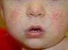
Rice. 2
Biochemical properties
Most bacteria of the Escherichia coli group (ECG) do not liquefy gelatin, coagulate milk, break down peptones with the formation of amines, ammonia, hydrogen sulfide, and have high enzymatic activity against lactose, glucose and other sugars, as well as alcohols. They do not have oxidase activity. According to the ability to break down lactose at a temperature of 37 ° C, BGKP are divided into lactose-negative and lactose-positive Escherichia coli (LCE), or coliforms, which are formed according to international standards. From the LKP group, fecal Escherichia coli (FEC) stand out, capable of fermenting lactose at a temperature of 44.5 ° C. These include E. coli, not growing on a citrate medium.
Stability in the external environment
E. coli are not heat tolerant. Bacteria of the Escherichia coli group are neutralized by conventional pasteurization methods (65 - 75 ° C). At 60 ° C, E. coli dies after 15 minutes. A 1% solution of phenol causes the death of the microbe after 5-15 minutes, sublimate at a dilution of 1: 1000 - after 2 minutes, resistant to the action of many aniline dyes. The persistence of Escherichia coli at low temperatures and in various environmental substrates has not been studied enough. According to some reports, E. coli can persist in water and soil for several months.
Sanitary-indicative value
The sanitary and indicative value of individual genera of bacteria of the group of Escherichia coli is not the same. The detection of bacteria of the genus Escherichia in food, water, soil, and equipment indicates fresh fecal contamination, which is of great sanitary and epidemiological significance. It is believed that bacteria of the genera Citrobacter and Enterobacter are indicators of older (several weeks) faecal contamination and therefore they have a lower sanitary value than bacteria of the genus Escherichia. With prolonged use of antibiotics, various variants of Escherichia coli are also found in the human intestine. Of particular interest are the lactose-negative variants of Escherichia coli. These are modified Escherichia that have lost the ability to ferment lactose. They are isolated from human intestinal infections (typhoid fever, dysentery, etc.) during the recovery period. E. coli that do not grow on Coser's medium (citrate medium) and ferment carbohydrates at 43-45 ° C (E. coli) have the greatest sanitary and indicative value. They are an indicator of fresh fecal contamination.
Diseases caused in humans by E. coli
Intestinal diseases caused by pathogenic E. coli are united under the general name of escherichiosis. The terms coli-infection, coli-enteritis, travelers' diarrhea, colibacillosis are also used.
Escherichiosis refers to acute intestinal diseases (AII) with a fecal-oral mechanism of infection. Each of the above classes of pathogenic E. coli is characterized by certain differences in the course of the disease, which in its symptoms may resemble cholera or dysentery. The incubation period lasts 3-6 days (usually 4-5 days).
Media and distribution
As already mentioned, E.Coli bacteria are part of the normal intestinal flora of not only humans, but also cattle and pigs. The young of the latter are often infected with colibacillosis and, accordingly, their meat (beef or pork) can serve as a source of infection. Pets (dogs, cats) are also susceptible to this disease, but the main way of infection is still fecal contamination of drinking water or food.
Human danger
The infective dose strongly depends on the type of pathogenic E. coli (for example, for enterotoxigenic E. coli, this value can be from 100 million to 10 billion bacteria, while for enteroinvasive and presumably enterohemorrhagic E. coli - only 10 organisms, like Shigella). The most susceptible to the disease are young children, the elderly and debilitated people.
In children, escherichiosis occurs in the form of varying severity of enteritis, enterocolitis in combination with a syndrome of general intoxication. In moderate and severe forms, it is accompanied by fever, diarrhea, sepsis.
In adults, the disease caused by E. coli resembles the course and clinical symptoms of acute dysentery. It occurs more often in erased and mild forms, less often (15-20%) there is a moderate and severe (3%) form.
The prognosis in adults and children older than a year is favorable, the most severe disease occurs in children in the first half of life.
Microbiology: lecture notes Tkachenko Ksenia Viktorovna
2. Escherichia
2. Escherichia
The genus Escherihia includes seven species. The most important species is E. coli, which are divided by pathogenicity into:
1) pathogenic (diarrheal);
2) conditionally pathogenic (they are part of the normal intestinal microflora).
They are mobile, do not form capsules.
Biochemical properties:
1) ferment glucose with the formation of acid and gas;
2) ferment lactate.
Antigenic structure:
1) according to the O-antigen, they are divided into serogroups (more than 160);
2) the majority have K-AG and N-AG.
Diseases caused by Escherichia are divided into two groups:
1) endogenous co-infections; are caused by their own Escherichia coli, which, with a decrease in immunological reactivity, causes purulent-inflammatory diseases;
2) exogenous coli infections - escherichiosis. These are typical intestinal infections, caused only by pathogenic E. coli that enter the body from outside. The main source is man.
Pathogenic E. coli are divided into four main classes.
1. ETEC - enterotoxigenic Escherichia coli. They have a tropism for the epithelium of the small intestine. Once in the body, they attach to the receptors of enterocyte membranes. They have the SF factor of colonization, due to which they populate the epithelial cells of the small intestine. They do not penetrate inside the cells, and inflammation does not develop.
They produce exoenterotoxin, the synthesis of which is encoded by the plasmid. This toxin is made up of:
1) LT-thermolabile fraction;
2) ST-thermostable fraction.
The toxin has a cytotonic effect. As a result of its impact, the process of enterosorption is disrupted, which leads to the development of diarrheal syndrome. Clinically, the disease proceeds as a mild form of cholera.
2. EIEC - Enteroinvasive Escherichia coli. They have a tropism for the epithelial cells of the large intestine. The factors of their virulence are the presence of outer membrane proteins on the surface of the cell wall, the ability to invade and intracellular reproduction. Bacterial reproduction leads to cell death. In place of dead cells, ulcers and erosion are formed, surrounded by inflammation.
3. EPEC - enteropathogenic Escherichia coli. Cause enterocolitis in children under one year old. The epithelium of the small intestine is affected. Virulence factor - the ability to limited invasion.
4. EHEC - enterohemorrhagic Escherichia coli. They have a tropism for the epithelial cells of the large intestine. The virulence factor is the production of two types of Shigo-like toxins (SLTs). Cause hemocolitis.
The main diagnostic method is bacteriological examination.
It is necessary to determine:
1) belonging of the isolated E. coli culture to the pathogenic serogroup (agglutination and precipitation reactions);
2) the presence of a toxin (using enzyme-linked immunosorbent assay (ELISA)), if the isolated structure belongs to the ETEC serogroup;
3) the presence of outer membrane proteins (ELISA), if the isolated structure belongs to the EIEC serogroup;