The share of gallbladder cancer accounts for about eight percent (and among oncological pathologies of the gastrointestinal tract, it is no more than 0.5%), which is why many general practitioners do not know the specifics of its detection and treatment tactics.
Most often, a malignant neoplasm develops from the cells of the mucous membranes of the bottom of the gallbladder or its neck.
Definition and statistics of cancer
Gallbladder cancer belongs to the category of rather rare malignant tumors that affect the tissues of this bean-shaped organ located at the bottom of the liver and designed to store and accumulate a special fluid - bile.
Produced by liver cells, bile is an indispensable participant in the digestive process.
Among tumors of the gallbladder, cancer ranks first, and in 74% of cases it is detected in patients who also have cholelithiasis or cholecystitis. Thus, any factor leading to the appearance of gallstones is capable of provoking the development of gallbladder cancer, since the disease tends to develop in a calcified gallbladder.
In the photo, an ultrasound diagnosis that shows gallbladder cancer
Women are four times more likely to experience it than men. As a rule, patients over the age of fifty are susceptible to this disease.
Causes and risk factors
The specific causes that are responsible for the development of gallbladder cancer are not known for certain, therefore, it is believed that the following risk factors most often contribute to the activation of the oncogene:
The presence of:
- long-term cholelithiasis (it is assumed that the impetus for dysplasia of epithelial tissues is their chronic inflammation and constant traumatization);
- sclerosing cholangitis (inflammation of the liver);
- adenomatous polyps of the gallbladder, the diameter of which exceeds one centimeter;
- chronic cholecystitis;
- and polycystic liver disease.
Types
The different histological structure of malignant neoplasms of the gallbladder is the basis for dividing it into different types, represented by:
- skyrr;
- poorly differentiated cancer;
- mucous cancer;
- solid cancer;
All types are characterized by a high degree of malignancy and a tendency to early metastasis (most often using the lymphatic tract).
The first symptoms of gallbladder cancer
In the early stages of the disease, there are practically no specific signs. As a rule, at this stage of development, gallbladder cancer is discovered quite by accident during a histological examination of tissues removed during a cholecystectomy operation for calculous cholecystitis.
A tenth of patients have migrating thrombophlebitis (the so-called Trousseau's syndrome). With this syndrome, phlebothrombosis is formed in different parts of the body, which is practically not treatable.
The early period of gallbladder cancer, characterized by the presence of non-specific manifestations, is often called pre-icteric. At this stage, patients have a feeling of bloating in the epigastric region, heaviness in the right hypochondrium, various stool disorders, frequent nausea, severe weakness, a state of constant malaise, sudden weight loss.
The duration of the preicteric period is due to the localization of the tumor focus and its proximity to the bile ducts. With the localization of the tumor process in the tail and body of the pancreas, the pre-icteric period lasts much longer than with the defeat of its head or extrahepatic ducts.
General symptoms of manifestations
 With the further development of a malignant neoplasm, intense obstructive jaundice develops, accompanied by a whole range of symptoms.
In a number of cases, they are the first to indicate the presence of a far-reaching process.
Jaundice is caused by the germination of the tumor or mechanical compression of the bile duct, which prevents the free outflow of bile into the duodenal cavity.
For the icteric period, in addition to persistent jaundice, a significant increase in the liver, the presence of nausea, vomiting, constant skin itching, a change in the color of urine (it darkens) and feces (it becomes lighter) are characteristic.
Blockage of the bile ducts by malignant neoplasms leads to empyema or dropsy of the gallbladder, inflammation of the bile ducts (cholangitis), and secondary biliary cirrhosis.
Damage to the liver by cancer cells leads to the appearance of symptoms of liver failure, which is expressed by lethargy, slowing down of mental reactions, severe muscle weakness (adynamia).
Advanced gallbladder cancer leads to peritoneal carcinomatosis, abdominal dropsy (ascites), and extreme malnutrition (cachexia).
Stages of the disease
- At the zero stage, mutated cells concentrated on the inner wall of the gallbladder begin to actively affect its healthy tissues.
- Stage 1 disease is characterized by the presence of a small elongated or oval neoplasm, localized on the wall of the gallbladder and slightly protruding into its cavity. Outwardly similar to a polyp, it is distinguished by the rapidity of its growth. The tumor of the first stage in its development goes through two stages. During the first, the walls of the gallbladder are damaged: its inner and connective tissue layers. During the second stage, the tumor captures the cells of the muscle tissue and another connective layer.
- The development of a stage 2 tumor is also characterized by two stages. The first is the defeat of the visceral peritoneum. Then the tumor process spreads to the tissues of the pancreas, liver, large and small intestines and nearby lymphatic vessels.
- At stage 3, a malignant neoplasm affects the blood vessels of the liver, gaining the ability to spread throughout the body.
- Stage 4 is characterized by distant metastasis and damage to distant organs and lymphatic vessels.
Ways of metastasis
Gallbladder cancer can metastasize in three ways:
- By sprouting into adjacent tissues (, pancreas, large and small intestines,).
- Lymphogenous route (through the lymphatic vessels).
- Hematogenous way (through the blood vessels along with the bloodstream).
Diagnostics
 The prolonged asymptomatic course, as well as the low specificity of its manifestations, are responsible for the fact that in most (70%) cases, gallbladder cancer is diagnosed already at the stage of an inoperable tumor.
- Physical examination of the patient reveals an increase in the gallbladder, spleen and liver, as well as the presence of an infiltrate in the abdominal cavity.
- To determine the operability of the tumor and the presence of metastases, diagnostic laparoscopy is performed.
- and allows not only to identify a number of pathological changes that have occurred in them as a result of the tumor process, but also to help with the collection of biomaterials during the puncture.
- If the diagnosis is in doubt, a percutaneous biopsy of the gallbladder is performed.
- In the patient's blood, the concentration of the cancer-embryonic antigen is measured and performed.
- Clarifying diagnosis is performed by methods, percutaneous transhepatic cholangiography, retrograde and cholescintigraphy.
- Treatment of gallbladder cancer should be radical. When diagnosing it in the early (0, I and II) stages, a simple or extended cholecystectomy (removal of the gallbladder) is performed.
- In stage III cancer, a more voluminous operation is performed, in addition to cholecystectomy, which also includes excision of the affected tissues of the right lobe of the liver. If indicated, the pancreas and duodenum are removed (pancreatoduodenectomy).
- With an inoperable tumor, a whole range of palliative measures are performed to reduce jaundice by recanalization (restoration of the lumen) of the bile ducts or by creating a new path for the outflow of bile by applying a superficial bile fistula.
Not all neoplasms that arise in the gallbladder manifest themselves immediately. It all depends on the location, histological structure and malignancy of the tumor.
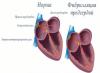
The structure and functional features of the gallbladder
The gallbladder is a pear-shaped hollow organ designed to accumulate and store bile produced by liver cells. Anatomically, it consists of the bottom, body and neck. Bile is excreted through the cystic duct, which is a continuation of the neck of the bladder, then into the common bile duct.
Organ topography
The gallbladder is located directly under the liver, more precisely under the right lobe. The gallbladder bed is made up of ligaments that attach it to the lower edge of the liver and hold it in a fixed position.
Regarding bone structures, the gallbladder is located in the right hypochondrium, 2 cm to the right of the midclavicular line.
Functions of the gallbladder and bile:
- reservoir and "storage" - bile accumulates for the next portion of food;
- participation in the processing of nutrients, in particular, the emulsification of fats;
- activation of pancreatic enzymes to accelerate the breakdown of carbohydrates, proteins and lipids in the gastrointestinal tract;
- synchronization of the pancreas - along with bile, pancreatic enzymes are secreted into the lumen of the duodenum. This contributes to timely and complete digestion;
- regulation of small intestine motility. The chemical receptors of the intestinal cells react to bile - the promotion of food and the absorption of nutrients begin.
A little about the incidence statistics
Neoplasms of the digestive system in the list of oncological diseases occupy approximately 6-7th place. But specifically, tumors of the gallbladder, bile ducts and liver are rare, it is no more than 1–1.5% of all oncopathologies.
The increase in incidence over the past 5 years is no more than 0.4%. Age limit of pathologies: people over 60 years old. Although liver tumors have become “younger” over the past decade, this is due to an increase in the incidence and its transition to hepatocellular carcinoma.
Modern medicine has in its arsenal accurate diagnostic devices, high-tech methods of biliary tract surgery, and targeted therapy. Often the symptoms of liver and gallbladder tumors are disguised as other, more common diseases: hepatitis, biliary cirrhosis.
Therefore, cancer of the biliary system is often diagnosed in the later stages, when the possibility of curing the disease is very small. In this case, it is necessary to do palliative (temporarily alleviating the patient's condition) operations.
Classification of tumors of the hepatobiliary system
Tumors are divided into several groups, depending on the sign of classification.
I. Histological classification:
Benign tumors and / or precancerous diseases:
- Hemangiomas and adenomas of the liver, true liver cysts.
- Fibroma, adenoma, myxoma, leiomyoma, fibroxanthogranuloma of the gallbladder, papillomas.
Malignant tumors:
- originating from the epithelium: adenocarcinoma (most common), solid and mucous cancer of the gallbladder with varying degrees of differentiation; hepatocellular carcinoma (hepatocellular carcinoma). The closer in structure to the normal tissue of the tumor, the easier they are to treat.
- Growing from the connective tissue layer: skirr (fibrous cancer) of the gallbladder and liver sarcoma.
- Growing from parenchymal structures: hepatoma (a tumor formed from liver cells), cholangioma (a tumor from the intrahepatic bile ducts), cholangiohepatoma;
- Anaplastic cancer is the most malignant tumor of the gallbladder. It grows rapidly and metastasizes, but is also rare.

II. Classification according to the degree of damage by atypical cells of organ structures:
- superficial process - affects only the mucous membrane;
- deep process - the tumor spreads into the thickness of the wall of the organ or "spreads", affecting more anatomical structures of the gallbladder;
- the tumor goes beyond the organ - cancer cells affect the liver, "local" lymph nodes, stomach or small intestine;
- total spread of the tumor - beyond the above localizations - by hematogenous and lymphogenous routes.
III. Alphanumeric classification according to the international TNM system, where T is the extent (depth) of the tumor, N is the presence/absence of metastases in regional lymph nodes, M is distant metastases.
Benign tumors of the gallbladder are rare. Usually they are an incidental finding on ultrasound of the abdominal organs or on radiopaque examination of the biliary tract. They are asymptomatic or asymptomatic. The most that can show up are symptoms. A person with such a pathology is under the supervision of a gastroenterologist: if the tumor does not grow and symptoms do not progress, then it is not even removed.
But malignant tumors in the gallbladder are more common than benign ones. They make up 90% of all neoplasms of the biliary tract.
Causes and risk factors for gallbladder tumors
Until now, the causes of cancer are unknown. Some experts consider genetic mutations to be “guilty” of the appearance of atypical cells, others - the way of life of a person. Any combination of several predisposing factors can be dangerous.

Risk factors for developing gallbladder cancer:
- heredity - if any of the family members had a history of cancer, then there is a likelihood of it occurring in the next generations;
- with exacerbations - inflammation provokes the formation of polyps, which tend to become malignant. Even a tumor in the form of a small polyp of the gallbladder can give unpredictable consequences;
- cholelithiasis - stones injure the wall of the bladder and ducts. At the site of damaged cells, atypical ones may form;
- a combination of 2 and 3 states - a long-term cholelithiasis, accompanied by symptoms of chronic bacterial cholecystitis. This is a particularly dangerous combination in terms of carcinogenesis;
- bile stasis caused by biliary dyskinesia of the hypokinetic type - occurs when the contractility of the duct wall is impaired;
- malnutrition and constant errors in the diet - an excess of fatty and carbohydrate foods disrupts the dynamics of the outflow of bile. And a small amount of plant food reduces intestinal motility and contributes to dyskinesia of the biliary tract;
- concomitant pathology of the upper gastrointestinal tract - chronic with reflux, pancreatitis, duodenal ulcer;
- harmful chemicals and heavy metals - long-term observations confirm that metallurgy workers are more susceptible to the occurrence of neoplasms in the gallbladder.
Symptoms of neoplasms of the gallbladder
All symptoms of tumors of the gallbladder and ducts can be divided into two groups: local and general.
- Local (local) symptoms - signs of the disease, manifested in the organ system where the tumor is localized.
- General symptoms are signs of a disease that affect the functioning of the body as a whole.
The first symptoms of a neoplasm of the biliary tract can be mistaken for biliary dyskinesia, which is present in 50% of the Russian population. Often, alarming "calls" from cancer are attributed to dietary errors, fatigue, overwork or gastritis, which also affects most of the country's inhabitants.

As the disease progresses, more serious manifestations “connect” to the clinical picture.
1 group of symptoms - local:
- pain in the right hypochondrium and / or in the epigastrium, tending to spread throughout the abdomen;
- bitterness in the mouth, which is associated with an obstruction to the outflow of bile;
- vomiting due to impaired motility of the biliary tract and intestines;
- bloating, flatulence due to the lack of adequate digestion of fats and impaired gastrointestinal motility;
- lightening of the stool (up to a pale yellow tint). Normally, due to the bile pigments oxidized during digestion, the feces become brown. If bile does not enter the intestine due to obstruction by the tumor, then the stool does not turn into the usual color.
Group 2 symptoms - general:
- signs of intoxication of the body - loss or perversion of appetite, nausea, weakness;
- yellowness of the skin and mucous membranes - bile constantly accumulates, but does not enter the intestine due to the blockage of the lumen of the biliary tract by the tumor. The "search" for an alternative output begins - absorption into the blood;
- fever - immunity with the help of fever tries to fight atypical cells.

Consequences and complications of tumors of the biliary system:
- - the tumor closes the lumen of the bile duct and interferes with the outflow of bile;
- biliary pancreatitis - the common bile and pancreatic ducts have one outlet. The output of bile and pancreatic juice occurs synchronously. If bile does not enter the intestinal lumen due to a tumor, the outflow of pancreatic juice with enzymes is delayed. Self-digestion of the pancreas begins;
- edema - local and generalized. They occur due to the "clamping" of the liver veins by an overgrown tumor - the pressure in the portal vein system increases, the venous outflow from the periphery is disturbed. Due to carcinomatosis (multiple metastases) of the peritoneum, ascites may occur - a lot of fluid in the abdomen.
Stages of gallbladder cancer and ways of metastasis
Stage 0- carcinoma is within the mucosa of the gallbladder.
Stage 1- the tumor has endophytic growth, i.e. it penetrates into the muscle and connective tissue layer of the organ wall.
Stage 2- defeat by cancer cells of the nearby digestive organ (liver, stomach, pancreas) and regional lymph nodes.
Stage 3- metastases enter other organ systems through the blood or lymphoid bed.
Stage 4- Multiple metastases and cancer cachexia (wasting).

The main and most common ways of metastasis of gallbladder cancer:
- mesenteric, gastric lymph nodes, lumbar lymphatic trunk, retroperitoneal lymph nodes;
- pancreas;
- liver;
- stomach;
- spleen;
- distant metastases - inguinal lymph nodes, secondary malignant neoplasms in the lungs.
Which doctor to contact
In oncopathologies of the biliary tract, 4 specialists work in tandem: a gastroenterologist, an oncologist, an abdominal surgeon and an endoscopist surgeon.
The gastroenterologist observes the patient throughout the entire period of the disease and prescribes conservative therapy. The oncologist and the abdominal surgeon plan the course of the operation and perform it. The endocopist applies modern methods of invasive diagnostics of pathologies of the biliary tract.
Diagnosis of gallbladder cancer
There are laboratory and instrumental research methods.
Laboratory diagnosis is nonspecific. It will show the presence of "disorder" in the hepatobiliary tract, but does not identify the cause.
A biochemical blood test for a tumor will show that bilirubin, hepatic transaminases, pancreatic amylase (if biliary pancreatitis has developed), thymol test are elevated; increased gamma-globulin fraction of proteins against the background of a decrease in total protein.

Coprogram - in the feces there are undigested fats and various dietary fibers.
Clinical analysis of blood - there are leukocytosis and anemia.
Detection of cancer antigens in venous blood - carcinoembryonic and CA 19–9.
Instrumental methods are aimed at identifying a specific pathology:
- ultrasound examination of the abdominal organs - gallbladder tumors are clearly visible on ultrasound;
- radiopaque examination of the biliary tract will show the exact location of tumor stenosis;
- MRI of the abdominal cavity shows layer-by-layer clear localization and features of the tumor;
- scintigraphy - a radiological study that assesses the structure of the tissue and the speed of excretion of the isotope;
- laparoscopy - an operation to introduce probes into the abdominal cavity through small holes with visualization of organs and tissues. During laparoscopy, a biopsy is often taken - pieces of tissue are cut off for histological examination under a microscope. The diagnosis of cancer is confirmed and its type is determined.
Treatment of gallbladder cancer
Treatment of gallbladder tumors is divided into two large subgroups: conservative and surgical.
Surgical treatment
Treatment is radical, when the tumor is removed completely by surgery, and palliative, if the tumor cannot be removed without damaging important body structures, therefore, an operation is performed that temporarily alleviates the patient's condition.

Radical operations:
- removal of the gallbladder (cholecystectomy) - laparoscopic method or open access;
- cholecystectomy with partial resection of the liver - done if the cancer cells have partially moved to the liver.
Palliative operations:
- stenting and expansion of the bile ducts with the installation of a mesh implant;
- creating a bypass anastomosis between the gallbladder and duodenum 12;
- cholecystostomy - removal of the drainage tube from the gallbladder to the outside.
Conservative treatment
In a complex or various combinations, several methods of conservative therapy are used:
- chemotherapy is the standard administration of chemotherapy drugs orally or by intravenous drip. It has a lot of side effects, but with multiple metastases it is indispensable;
- radiation therapy - targeted irradiation occurs at the site of the affected organ;
- Targeted therapy for gallbladder tumors is considered more effective and safer than the 2 previous methods. It consists in the targeted effect of the medicinal substance specifically on cancer cells. This helps to minimize side effects and accelerate the destruction of the tumor;
- hepatoprotectors, antispasmodics, prokinetics - correction of the biliary tract and intestines.

During treatment, you must adhere to a diet: exclude fatty and fried, easily digestible carbohydrates; eat more boiled and stewed vegetables.
Forecast
If the tumor is detected in a timely manner, then with its complete removal, the outcome is favorable and a 5-year survival after surgery is ensured. Also, the prognosis of the disease is determined by the following factors:
- the prevalence of the process - in the early stages it is much easier to radically remove the neoplasm than in the later ones;
- histological type of tumor - if the cancer is highly differentiated, the chances of getting rid of it increase significantly;
- the consequences that the gallbladder tumor gave to the body - prolonged jaundice contributes to severe intoxication, metastasis of cancer cells - the forced use of toxic chemotherapy drugs;
- the possibility of radical removal of a tumor of the gallbladder.
A correct and timely diagnosis is the key to a complete and successful cure. A modern method of treatment - targeted therapy - for tumors of the gallbladder increases the survival of patients.
Cancer is curable. But you need to listen to your feelings and visit a gastroenterologist at least once every two years.
Gallbladder cancer accounts for 20% of all oncological diseases of the gastrointestinal tract (GIT), and in most patients it is the result of a long course of gallstone disease (GSD). According to statistics, the development of benign formations in the gallbladder (GB) is much more common, and pathological tumors are observed in 0.5–4% of patients due to prolonged cholelithiasis.
The main risk group includes women over 60 years of age who have repeatedly given birth and are overweight. Among them, the risk of morbidity is 2–5 times higher than among men. It is believed that the process of progression of severe dysplasia and its degeneration into gallbladder cancer occurs within 15 years. However, the disease can develop faster if the following risk factors are present:
- excess weight - metabolic disorders and a significant predominance of carbohydrates over fiber in the daily diet significantly increases the risk of gallbladder cancer;
- heredity - 13.6% of patients are characterized by a family history of oncology of the gastrointestinal tract;
- harmful work - there was an increased risk of developing tumors of the hepatobiliary system among workers in the chemical and metallurgical industries due to inhalation of nitrosamine vapors and other poisons;
- infections of the digestive tract - chronic bacterial invasions increase the risk of developing gallbladder cancer several times, for example, typhoid fever - 6 times;
- pathological neoplasms - emerging benign tumors of the gallbladder can self-destruct if a person adheres to the doctor's recommendations, or they can degenerate into cancer;
- bad habits - alcohol disrupts the process of bile production by the liver, which leads to cholelithiasis, and nicotine and tobacco tar have a mutagenic effect on the cells of the hepatobiliary system, contributing to their degeneration.
The presence is not a harbinger of cancer, since it can occur in the absence of stones. Statistics say only that 3 out of 4 people with gallbladder cancer have stones.

After diagnosing cholelithiasis, the question of treatment arises: conservative or surgical? If small cholesterol formations were found in the hollow organ, then drug treatment can be dispensed with. Calcium deposits are of great danger, since they are difficult to "dissolve" with drugs and remove from the body. In addition, calcium begins to be deposited in the layers between the muscular, serous and mucous layers of the gallbladder wall, where the gallbladder tumor most often begins to form.
Doctors advise resection of a hollow organ for patients with polyps larger than 10 mm, as they often degenerate into oncology. The presence of a cyst in the common bile duct is also an alarming sign, since it may contain areas of precancerous changes. In the case of an increase in the cyst, resection of the organ is indicated for the prevention of oncology.
In the US, where fast food is a daily staple for most of the population, gallbladder cancer is three times more common than in other countries. This suggests the significant role of malnutrition as a pathogenic factor in the development of oncology of the gastrointestinal tract.
Symptoms and stages
Three-quarters of patients with gallbladder cancer are over 70 years of age, as the disease is most often diagnosed at an advanced stage. This is due to the absence of nerve receptors in the hollow organ and, as a result, its "silence".

The first manifestations of the disease are nonspecific: general weakness and fatigue, periodic, barely noticeable dull. Such signs can easily be attributed to stress, fatigue and overeating. As the disease progresses, the person begins to feel worse, and the basis of the clinical picture is the signs of acute cholecystitis: bloating, nausea, vomiting, pain in the right side, sudden weight loss and jaundice. In the elderly, a typical symptom of gallbladder cancer is such a clinical duet as persistent dull pain and low-grade fever.
With tumors of the bile ducts, jaundice is observed in 96–100% of patients, and with oncology of the gallbladder, this symptom manifests itself only in 57% of patients.
Doctors classify the disease in different ways: according to morphological changes, according to the size and structure of oncology, according to the specifics of the results of histological studies, etc. But the main thing is the division according to the predominant clinical syndrome and the stages of progression of the pathology. In the first case, the following forms are distinguished: "tumor", dyspeptic, icteric, septic and metastatic or "silent".
In the second case, all patients are divided into two large groups: localized cancer (stage I) and non-localized (II–IV). Already at the second stage, the tumor grows through the wall of the gallbladder into the ducts, due to which one resection of the organ becomes insufficient for effective treatment. Therefore, other methods are used: chemotherapy, radiation exposure, occlusion of blood vessels that "feed" the tumor, etc. However, there is an exception in this group - these are patients in whom the oncoprocess develops only in the lymph nodes. In this case, the surgical operation does not cause serious difficulties.
The stages of the pathological process are characterized by such structural changes:
| Stage | Changes | |
|---|---|---|
| 0 | – | the presence of abnormal cells in the layers of the wall of the gallbladder; |
| I | A | the formed tumor grows through the mucous layer; |
| B | the tumor grows through the muscle layer; | |
| II | A | the spread of the tumor to the peritoneum covering the gallbladder, and the connective tissues of neighboring organs; |
| B | spread of the tumor to nearby lymph nodes and muscle layers of neighboring organs; | |
| III | A | transition of the tumor to the neighboring organ through the visceral peritoneum; |
| B | damage to blood vessels and lymph nodes of nearby organs; | |
| IV | A | spread of the tumor to the main artery of a neighboring organ; |
| B | spread of the tumor to the lymph nodes along the large arteries. |
Progression of oncology
Gallstone disease can be a harbinger of cancer for decades without triggering the growth mechanism of a gallbladder tumor. If abnormal cells appear, then they begin to spread very quickly, passing through such generalization paths:
- Direct damage to the liver tissues (occurs in 58–91% of patients) - in this case, operable treatment is still possible with the removal of the IV and V segments of the liver.
- Metastasis (hematogenous and lymphogenous) - such a lesion is observed in 68% of those who died from cancer of the gallbladder at autopsy. Metastases are found in 94% of asymptomatic patients with oncology. Most often they affect the liver (50–85%), and the lungs are in second place (5–34%), therefore, with cancer of the gallbladder, a chest x-ray is necessary to detect metastases.
- Peritoneal metastasis - detection of abnormal cells in the peritoneum occurs in 60% of cases.
Diagnostics
With modern medical equipment, ultrasound has ceased to be the main instrument for examining internal organs. Its sensitivity for detecting gallbladder cancer is only 29% and its diagnostic accuracy is 62%. Therefore, it is rather an assistant for a more accurate diagnosis. For example, contrast and endoscopic ultrasound can differentiate malignant tumors from benign ones with an accuracy of 89%, while intraoperative ultrasound provides 100% accurate data on the nature and stage of gallbladder cancer.

Since ultrasound diagnostics remains the most affordable in financial terms, it is with it that the examination of most patients begins. If an unresectable tumor has been identified, the doctor draws up a therapeutic regimen based on the results of instrumental and laboratory studies. If gallbladder cancer is detected at an early stage or the data obtained are inaccurate, CT or MRI is performed to more clearly visualize structural changes in the organ. But the study of the structure of the tumor at the micro level is carried out through a biopsy. Along with an instrumental examination, laboratory tests are prescribed to determine the quality of the functioning of the liver and identify cancer markers.
Treatment
The main therapy for gallbladder cancer remains tumor resection, which involves the removal of a hollow organ, as well as parts of the liver and urinary tract, if cancer has spread to them. With localized cancer, the survival rate is quite high (70-80%), but every second patient relapses within a year with the spread of abnormal cells to the biliary tract and adjacent organs of the gastrointestinal tract, most often the liver, pancreas and duodenum. If the localization of the tumor is not limited to the gallbladder, then the survival rate after surgery for a year is no more than 20%.

Chemotherapy is prescribed as an additional therapy that allows to delay the operation until the patient's condition stabilizes and the severity of symptoms of gallbladder cancer decreases. It increases the patient's survival by 6–9 months, but cannot destroy all abnormal cells. Cryodestruction is also ineffective. And since there is no treatment for oncology of the gallbladder yet, the best way is to prevent cholecystitis, cholelithiasis, and, consequently, cancer.
Forecast
The average death rate for gallbladder cancer is 19.6%, but the real figure is much higher. Due to the impossibility of early diagnosis, 70% of patients die in the next two years after diagnosis and surgery due to recurrence of oncology in the bile ducts and liver. If the disease was detected at stage IA, then the five-year survival rate is 95–100%.
Recent studies have shown that in stages IB-IIIA, survival is significantly increased when performing surgery with resection of the gallbladder and affected segments of the liver. Radiation and chemotherapy are ineffective and only for a few months relieve the symptoms of gallbladder cancer. Therefore, such treatment is used more to prepare the patient for a major operation.
High mortality in this oncological disease is also due to the advanced age of the majority of patients who have a number of contraindications to surgical intervention. They also have a high risk of developing tumor intoxication and other complications that increase the likelihood of death. In the case of pancreatoduodenal resection, 90% of patients develop metastases in the first year after surgery. Thus, both surgical and conservative therapy for stage II–IV cancer is effective only for 5–12% of patients.
The gallbladder is a small, hollow, pear-shaped organ located under the liver. It concentrates and stores bile, the fluid needed to digest fats in the small intestine. With gallbladder cancer, it is important to start treatment on time, as the disease can be asymptomatic and often detected too late. Cancer can affect nearby organs and tissues and it is much more difficult to cure the disease.
Malignant tumors of the gallbladder are quite rare, more often develop in women, in 7 out of 10 cases.
Our company, the medical service "site", offers services for organizing the diagnosis and treatment of gallbladder cancer in Israel:
1. Selection of doctors and clinics, individual approach, taking into account all wishes, comfortable conditions of stay.
2. Carrying out all procedures as soon as possible.
3. Moderate prices due to contracts with medical centers for the provision of medical care at wholesale prices.
4. Providing a "second opinion" (obtaining advice from another specialist), after the end of treatment, maintaining contact with the doctor.
To get a consultation
Causes of gallbladder cancer
There are several risk factors associated with this type of cancer.
- Like most cancers, gallbladder cancer is more common in older people.
- A family history of gallbladder cancer increases the risk of the disease by 5 times, especially if there is a genetic mutation known as BRCA2.
- Risk factors are gallstones and inflammation (cholecystitis). 8 out of 10 people with gallbladder cancer are diagnosed with these disorders. Studies have shown that having a family history of stones increases the risk of cancer.
- "Porcelain" gallbladder - calcium deposition on the inner wall of the organ - one of the potential factors.
- Primary sclerosing cholangitis (PSC), a type of inflammation of the bile ducts, can increase the chance of cancer.
- Smoking and some industrial chemicals contain nitrosamines, chemicals that damage DNA and increase the risk of cancer.
- Some abnormalities of the pancreas and bile ducts increase the likelihood of this disease. These are congenital disorders - growths along the bile duct, an abnormal connection between the bile ducts and the pancreas.
- Gallbladder polyps are small growths that appear on the mucous membrane of the organ. The larger the polyp, the higher the risk that it will turn into a malignant one.
- Being overweight increases the risk of many types of cancer, including gallbladder cancer. Obesity means exceeding by more than 40% the maximum desired weight according to a certain height. Excess weight causes a change in the hormonal balance in the body, which increases the risk of this type of cancer.
- Diabetes increases the chance of gallbladder cancer and bile duct tumors, according to a number of studies.
- There is some evidence that diet affects the risk of developing this disease.
- Ethnicity is one of the factors. The highest incidence of gallbladder cancer is in North India.
- Salmonella infection increases the risk of this disease. According to some studies, Helicobacter pylori bacteria may also be affected.
Signs and symptoms of gallbladder cancer
In the early stages, the disease does not cause any manifestations. When the disease is diagnosed, it often goes beyond the primary focus and affects the surrounding tissues and organs.
Most of the symptoms are characteristic for the later stages of the development of the disease. It is worth knowing that other pathologies can also cause these symptoms.
- Abdominal pain may occur on the right side of the abdomen. Some people describe it as pulling. If cancer or stones block the bile duct, the pain will be acute.
- Nausea is very common in the advanced stages of gallbladder cancer. It is easily controlled with antiemetic drugs.
- Jaundice means that the liver is not functioning properly. Symptoms include: yellowing of the skin and whites of the eyes; dark urine; white stool. Jaundice is associated with the accumulation of bile salts in the blood. Half of people diagnosed with gallbladder cancer have jaundice, a sign of advanced cancer. It is worth remembering that hepatitis is a more common cause of jaundice.
- The expansion of the gallbladder occurs due to blockage of the bile duct and filling the organ with bile.
- Other less common symptoms include loss of appetite, weight loss, and constipation.
Types of gallbladder cancer
In 85 out of 100 cases of this disease, adenocarcinoma is diagnosed. This type of cancer develops in the mucus-producing glandular cells of the organ. Andenocarcinoma is divided into papillary, non-papillary and colloid.
Less common types of gallbladder cancer include squamous cell, small cell, and sarcoma.
Rare types of gallbladder cancer include neuroendocrine tumors, lymphomas, and melanoma.
Find out prices for treatment
Diagnosis of gallbladder cancer in Israel
 The oncologist collects an anamnesis, interrogates about the symptoms. Examines the patient, probing the abdomen for signs of enlargement. Checks the whites of the eyes and skin color for the symptom of yellowing. Examines the lymph nodes in the neck and groin.
Depending on the results, the following types of examinations can be assigned:
- Blood tests called liver tests. This is a series of tests that check the functioning of the liver and gallbladder. Also included is a test for bilirubin, a chemical in bile. A small amount of bilirubin in the blood is completely normal. But a high level usually means there is a problem with the gallbladder or liver.
- Ultrasound. If the tumor is found in the gallbladder, ultrasound can be used to determine whether the cancer has spread to the walls of the organ.
- A CT scan will show tumor growth in and around the gallbladder, whether the common bile duct, lymph nodes, or liver are involved.
If the scan shows abnormal areas around or inside the gallbladder, the following may be done:
- ERCP - X-ray of an organ using an endoscope (endoscopic retrograde cholangiopancreatography - ERCP). The patient swallows a flexible tube with which the doctor examines the inside of the small intestine and takes a biopsy of areas that look abnormal. This test shows the narrowing or blockage of the bile ducts of the pancreas, it helps to plan the operation. Takes from 30 minutes to 2 hours.
- MRCP is a type of MRI of the gallbladder, pancreas, and bile ducts. MRCP stands for magnetic resonance cholangiopancreatography. The procedure requires preparation, in 2 hours it is necessary to stop eating and drinking. MRCP is less uncomfortable than ERCP, does not require painkillers or other drugs, but does not allow tissue sampling.
- Biopsy and fine needle aspiration. A biopsy means taking a sample of tissue and examining it under a microscope. This is the only way to determine if a tumor is cancerous. But if the doctor is absolutely sure, based on the results of other tests, that it is cancer, a biopsy is not needed. The gallbladder will be removed anyway.
If a biopsy is needed, it can be done in a number of ways: during laparoscopy with ERCP or with a fine needle aspiration biopsy.
To monitor and further treat gallbladder cancer, as well as to perform an aspiration biopsy, the doctor uses CT or ultrasound to guide the needle to the right place. Takes a sample of cells and sends it to the laboratory for further study. A tissue sample may be taken from the liver or lymph nodes to see if cancer has spread to them. After a gallbladder biopsy, the patient stays in the clinic for several hours or overnight. This is necessary because there is a risk of bleeding.
If the tests indicate gallbladder cancer, further investigations may be needed to determine the extent of the cancer. Most often, cancer affects the liver - in 8 out of 10 people. It can also enter the lymph nodes in the abdomen. For the treatment of gallbladder cancer, the following types of diagnostics can be carried out.
- MRI shows soft tissue more accurately than CT. The use of MRI with cholangiography can show blockage of the flow of bile by the tumor, as well as the spread of cancer to the portal vein. If there is a metal in the body (for example, a pacemaker), this test is contraindicated.
- Endoscopic ultrasound uses an ultrasound scanner and an endoscope to help determine the stage of the cancer, whether the tumor has grown into the wall of an organ, or spread to the liver. All this facilitates the process of planning the operation.
- Cholangiography examines the bile ducts using dye, X-rays, and an endoscope. The procedure lasts 30-60 minutes, with its help you can find out if there is a tumor in the gallbladder, if the duct is blocked. If there is a blockage, a stent may be placed.
Laparoscopy is a minor operation. A laparoscope equipped with a camera and light is inserted into the abdominal cavity through small incisions and examined for signs of cancer. With the help of a laparoscope, the surgeon has the opportunity to look inside the gallbladder. Laparoscopy helps in planning surgery and choosing other cancer treatments for gallbladder cancer. The procedure requires general anesthesia and overnight hospitalization. During its implementation, a biopsy may be performed. If there are stones or an inflammatory process in the gallbladder, the surgeon will immediately remove the organ. This operation is called a cholecystectomy. The advantage of this type of treatment is a shorter recovery period.
Request a call back
Stages of gallbladder cancer
Stages indicate the growth and spread of the malignant process. Determining the stage of the disease is necessary to select the optimal treatment option for gallbladder cancer.
The TNM system is used to classify gallbladder cancer.
- T - indicates the size and spread of the gallbladder tumor.
- N - for the defeat of the lymph nodes.
- M - for the penetration of the tumor process into other parts of the body.
According to this classification, 5 stages are distinguished - T1 - T4 and very early - called Tis or carcinoma in situ.
Tis (cancer in situ) - the tumor is located inside the organ. At this stage, the disease is rarely diagnosed. More often occurs when the gallbladder is removed for other reasons, such as due to the presence of stones.
- T1 tumor began to grow into the wall of the gallbladder. The stage is divided into T1a and T1b. T1a indicates damage to the connective layer under the inner shell of the organ wall, T1b - about the penetration of cancer into the muscle layer located behind the connective.
- The T2 tumor is located in the gallbladder but has grown through the muscle layer into the next layer of connective tissue.
- T3 tumor has spread beyond the boundaries of the organ, spread to the liver or another nearby organ - the stomach, intestines or pancreas.
- T4 - cancer has invaded the portal vein or hepatic artery, has given secondary foci to two or more organs outside the liver.
There are three stages of damage to the lymph nodes in malignant tumors of the gallbladder
- N0 - lymph nodes are healthy.
- N1 - the tumor process has affected one or more neighboring lymph nodes, for example, along the bile duct or the main arteries of the liver.
- N 2 - pathological cells have spread to the lymph nodes located beyond the gallbladder.
M indicates the penetration of the tumor process into other organs and tissues.
- M0 - the malignant process did not affect distant organs or structures.
- M1 - secondary foci arose in other organs, for example, in the brain or lungs.
The combination of T, N and M gives a complete description of the stage of the disease.
Ask a Question
 Gallbladder cancer stages according to another classification
There are 4 main stages, some doctors also talk about stage 0.
Stage 0 or carcinoma in situ. This cancer is at an early stage. Malignant cells are found only in the tissue layer lining the gallbladder. There is a low risk of spreading the disease.
Stage 1. The earliest stage of invasive cancer. It means that the malignant process is located only in the inner layers lining the tissues of the gallbladder. No penetration into nearby tissues or organs. Stage 1 is identical to T1, N0, M0 according to the TNM classification.
Stage 2. Cancer grows through the muscle layer of the gallbladder wall into the connective tissue that follows it, remains localized within the organ. Stage 2 corresponds to T2, N0, M0 according to TNM.
Stage 3 is divided into 3A and 3B.
- 3A - cancer has grown through the walls of the gallbladder, there are no malignant cells in the lymph nodes. Corresponds to 3, N0, M0.
- 3B - the tumor is located within the boundaries of the gallbladder or has grown through the outer layer and struck the nearest lymph nodes. Identical to T1, T2 or T3, N1 or M0.
Stage 4 indicates metastatic cancer, divided into 4A and 4B.
4A - the tumor process has affected the artery leading to the liver, or has spread to 2 or more organs outside the liver. Adjacent lymph nodes (T4, N0 or N1, M0) may be affected.
4B indicates cancer of any size that:
- affected lymph nodes located beyond the gallbladder, but did not penetrate into distant organs (any T, N2, M0);
- has metastasized to structures or organs farther from the gallbladder (any T, any N, M1).
Simple staging system
Sometimes doctors use a simplified staging system to decide what treatment for gallbladder cancer is needed. There are three stages:
- Localized gallbladder cancer (1 and 2) - the tumor is located within the boundaries of the gallbladder, can be removed surgically.
- Inoperable gallbladder cancer (3 and 4) - the malignant process has spread beyond the primary tumor and cannot be removed surgically. Sometimes it is possible to resect a stage 3 tumor.
- Relapse - the disease returned after therapy. A secondary tumor may appear in the gallbladder or in another area.
Treatment prognosis for gallbladder cancer
The data of international statistics intended for general guidance are presented.
Unfortunately, the overall prognosis for gallbladder cancer is not very good. According to most medical articles, only 10 out of 100 people (10%) will live 5 years or more (5-10 survival). It mainly depends on the stage of the disease. Diagnosis is often made late, when the disease cannot be cured.
Perspectives by stage - 5-year survival or more after diagnosis.
- Stage 0 - for 80 out of 100 people (80%).
- Stage 1 - for 50 out of 100 (50%). Some doctors believe that removing nearby lymph nodes and some liver tissue during surgery will prevent recurrence - an extended cholecystectomy is performed.
- Stage 2 - for 25 out of 100 (25%).
- Stage 3 and 4 - less than 10 out of 100 (10%).
Reliability of static data
There is no static information that will tell you exactly what will happen. Each disease is unique, just like the human body. For example, the same type of cancer can develop at different rates in different people. Many individual factors can affect treatment and prognosis, and it is important to be aware of this.
Research results show that participation in clinical trials may improve prognosis.
Apply for treatment
In the list of diseases of the digestive system, doctors also call gallbladder cancer. Despite the rarity of this disease (only 20% of cases from cancerous tumors of the entire digestive system), this diagnosis is terrible with long treatment and the absence of symptoms at an early stage.
People who have already encountered such a diagnosis will certainly have numerous questions. How to identify oncology in the early stages? How long do people live with stage 4 gallbladder cancer? Is it possible to completely get rid of the disease? These issues are extremely important, so all aspects of diagnosis and treatment should be sorted out in order.
Basic concepts
What exactly is the gallbladder? This is a fairly small bean-shaped organ. It is located in the lower part of the liver. The main function of the gallbladder is to store bile, a special secretory fluid that is involved in the digestion of food.
Gallbladder cancer is an oncological disease. It is characterized by the appearance of pathological cells in the tissues of the organ. Over time, these cells begin to grow and divide, forming a tumor. Such a neoplasm blocks the proper functioning of the gallbladder and neighboring organs. The code for the international classification of diseases of gallbladder cancer (ICD-10) is C23.
It has been noticed that the female half of humanity is more susceptible to this disease: according to statistics, there are almost twice as many women with this diagnosis as men. Thus, in 2013 in Russia, tumors of the extrahepatic biliary tract were detected in 2180 women and 1122 men (separate data on the gallbladder are not available).
With regard to age categories, the majority of patients are people who have reached 50 years of age. Although doctors note: over the past decade, gallbladder cancer has been increasingly diagnosed in people 30 years and older. Cases of illness in children have also been identified, but they are isolated.
What is the difficulty of diagnosis and treatment? The main reason is the treatment of patients mainly in the last stages of the disease. This greatly complicates the treatment.
Reasons for the development of gallbladder cancer
Name the specific reasons that become the impetus for the development of atypical cells, scientists can not. However, constant statistics have revealed factors that may increase the risk of gallbladder cancer:
- These are various diseases of the gallbladder of an inflammatory nature, the presence of stones. 85% of patients with oncology of this type in the past had problems with the gallbladder. These are chronic inflammations of the organ, and stones. At the same time, it was noticed: the larger the size of the stones in the gallbladder, the higher the risk of a malignant tumor.

- Constant contact with certain substances. Among the patients there are many workers in hazardous industries (rubber or metallurgical industry). This is due to the high concentration of chemicals.
- Bile duct cyst. Such a pathological phenomenon is often called precancerous. The fact is that a cyst is a neoplasm filled with bile. Under certain conditions, the cyst can increase in size, and then degenerate into a malignant tumor and show symptoms of gallbladder cancer. At the first suspicion of a cyst, you should go to the clinic as soon as possible.
- "Porcelain" gallbladder. This medical term is used to define the pathological condition of the organ, in which all the walls of the gallbladder are covered with calcium deposits. This condition occurs with severe inflammation. Traditionally, the affected organ is removed, as it often causes oncology.
- Typhoid fever. To date, infection with typhoid fever is an extremely rare phenomenon, but if it occurs in a patient, the risk of developing signs of gallbladder cancer increases by almost 6 times.
- Age changes. In the body of absolutely every person with age, irreversible phenomena occur at the cellular level, which can provoke the growth of atypical cells. This is fully confirmed by statistics: most of the patients belong to the category of elderly people.
- Bad habits. The list can include smoking, excessive drinking of alcoholic beverages, unhealthy diet.
Tumor histology
Gallbladder cancer is usually divided into several categories, taking into account certain characteristics.
According to the histological structure of cells, several types of tumors are distinguished:
- squamous cell carcinoma - a tumor that occurs in the epithelial layer and mucous membrane;
- adenocarcinoma - such a neoplasm appears from the glandular cells located in the epithelium of the organ;
- scirrhous;
- solid - from the Latin word solidum (solid), such a tumor is a group of cells arranged in plates;
- poorly differentiated - cells of such cancer often have irregularly shaped nuclei and an abnormal structure.
Tumor localization
According to the location of the malignant neoplasm, 2 types of gallbladder cancer are distinguished:

Metastases are the spread of malignant cells from the primary focus (in this case, from the gallbladder) to various other tissues and organs of the human body. Most often, metastases of gallbladder cancer spread to the lymphatic system, liver, intestines, and stomach.
Stages of a malignant neoplasm of the gallbladder
For a more convenient classification and description of the pathological processes occurring in the human body, it is customary to differentiate gallbladder cancer at the stage:

- 0 stage- it is often called precancerous. At this time, pathological cells are located on the mucous membrane of the organ, and the size of the tumor is quite small. Starting treatment at stage 0 allows you to completely get rid of the disease, but it is extremely difficult to diagnose such oncology - there are no symptoms at all.
- 1 stage. Malignant cells penetrate not only into the mucous membrane, but also into the adjacent layers of tissues. The diameter of the tumor also grows. At this stage, the first symptoms of gallbladder cancer may appear, but they are practically not noticeable. In most cases, the detection of the disease at this stage occurs during a medical examination prescribed for other reasons.
- Stage 2 (moderate severity). This stage includes the period of active tumor growth. By this time, the neoplasm reaches an impressive size, but does not go beyond the gallbladder. The symptoms are getting worse.
- 3 stage. It is at this stage of tumor development that many patients turn to the clinic, as pronounced persistent symptoms appear. By this time, the tumor has already given near metastases.
- 4 stage. Gallbladder cancer at this stage has several characteristics at once. These are the large size of the tumor, damage to nearby tissues (that is, metastases to other organs), the presence of a large number of symptoms of the disease, and the low susceptibility of the tumor to treatment.
Clinical picture
The main thing that distinguishes cancer from many others is the complete absence of symptoms in the early stages. This is the main problem that explains the late visit of many patients to the doctor.

In addition, many of the symptoms of gallbladder cancer are very similar to those of some other non-cancer diseases (for example, chronic cholecystitis). At the same time, the manifestation of all symptoms is not at all necessary - they can vary depending on the type of cancer and its localization.
Among the first symptoms of gallbladder cancer appear:
- pain in the right side of the abdomen under the ribs (at first, pain appears quite rarely and is of a short-term nature, but intensifies as the tumor grows);
- bloating and feeling of heaviness;
- the appearance of frequent bouts of nausea, vomiting is possible;
- stool disorders (flatulence can suddenly be replaced by constipation);
- lack of appetite or its significant decrease.
If at this stage the person does not go to the doctor and treatment is not started, the tumor continues to progress. A little later, symptoms of gallbladder cancer appear, such as:
- pain in the right precostal space becomes more frequent and longer, they can radiate throughout the abdomen, back, neck or shoulder;
- severe nausea ends with vomiting, but even this does not bring relief;
- tumor growth leads to an increase in the size of the gallbladder - as a result of this, the enlarged liver can be felt on its own;
- a slightly yellowish tint of the skin appears;
- there is burning and itching of the skin;
- there is shortness of breath (not only after physical exertion, but even at rest);
- appetite may be good or absent altogether, while body weight is sharply reduced;
- long-term preservation of high body temperature (from 37 to 39 degrees);
- fatigue, feeling of weakness, apathy.
Another characteristic sign may be a change in the color of urine and feces. Urine becomes darker, and feces, on the contrary, lighter.
Initial examination of patients
The prolonged absence of symptoms at stage 1 of gallbladder cancer leads to the fact that in 70% of cases, patients go to the clinic when the tumor has already reached a significant size and requires long-term complex treatment.
To prescribe the most effective course of therapy, the doctor needs to get a complete picture of the disease. To do this, he prescribes a number of tests, and also conducts:
- Complete examination of the patient. At the initial appointment, the doctor needs to get as much information as possible from the patient's words. This will allow you to judge the intensity of the symptoms. Based on this, the severity of the current illness can be assumed.
- Acquaintance with the features of the life of the patient and the history of his illness. Such details allow us to judge the magnitude of the risk of developing cancer.
- Physical examination. This concept includes examination of the patient, measurement of body temperature, palpation of the liver area (for an increase in the size of the organ), examination of the skin and eye sclera for the presence of a yellowish tint.
Laboratory research
Conducting laboratory tests will not reveal gallbladder cancer, but the results of the tests will clearly indicate the pathological condition of a particular organ.

The following analyzes are carried out:
- General urine analysis.
- Fecal analysis (coprogram).
- Biochemistry of blood. In diseases of the gallbladder, an increase in the level of transaminases, bilirubin, and alkaline phosphatase is observed.
- Assign a blood test to identify tumor markers. Such diagnostics allows obtaining data on the presence of malignant cells in the body.
Instrumental diagnostics
Instrumental research methods can be safely called the basis of diagnosis, since it is from the results of these studies that the doctor receives information about the state of the gallbladder, the presence or absence of a tumor, its localization, size and presence of metastases:

The main treatment for this disease is surgery. During it, the surgeon removes the gallbladder. In this case, 2 options are possible:
- Cholecystectomy. A surgical operation in which the gallbladder is removed. This approach to treatment is possible only in cases of early detection of oncology.
- Cholecystectomy + At stage 3, the removal of the gallbladder will be ineffective, since the malignant cells have already spread to the liver tissue. In this case, during the operation, the right lobe of the liver is also removed. In some cases, resection of nearby lymph nodes will be required.
In the last stages of the disease, gallbladder oncology is considered inoperable, so surgery is not prescribed. This is explained by numerous metastases affecting the lymphatic system, liver, lungs, and brain. In this case, courses of radio- and chemotherapy are prescribed as treatment.
Radiotherapy is a method of cancer treatment in which the patient is exposed to ionizing radiation. The essence of the method is that malignant cells are sensitive to radiation, therefore, under such exposure, they are destroyed. Quite often, radiotherapy is also used as an additional effect before or after a surgical operation. This treatment is quite effective, but has severe side effects.

Chemotherapy is another way to influence the tumor without the use of a scalpel. In this case, the treatment is based on taking strong drugs that have a detrimental effect on pathological tumor cells. Depending on the stage, concomitant diseases and the general condition of the patient, the doctor prescribes an intravenous infusion of drugs or oral administration. Dosage and duration are strictly controlled by the attending physician. The entire treatment period is divided into courses with a break of several weeks.
Special diet for gallbladder cancer
Oncological diseases are a rather difficult test for the entire human body. At the same time, it is extremely important that the gallbladder is involved in digestion, and therefore nutrition issues during this period should be taken very seriously.
The diet of a cancer patient should be built in such a way as to relieve the gallbladder and liver as much as possible.
Meals should be at least 5-6 per day, and portions are made small.
You need to give preference to dishes with fiber and protein, which are easily digested.
You need to completely abandon heavy food: fatty, salty, fried, smoked, sweet.
The diet should be so varied that it includes vegetables and fruits, lean meats, fish.
Be sure to take a complex of vitamins prescribed by a doctor. Such a supplement to the diet will help restore human immunity.
Forecast
Each patient with such a diagnosis certainly wondered how long they live with gallbladder cancer. In fact, no one can give an accurate forecast. The result of treatment depends on several factors at once, namely: the stage of the disease, the age of the cancer patient, concomitant diseases, the type and location of the tumor.
At stage 1, more than 60% of patients can be cured of oncology.
Started treatment at stage 2 gives a five-year survival rate of patients in 30% of cases.
At stage 3, five-year survival is observed in 10% of cases.
The smallest cure rate for stage 4 gallbladder cancer is less than 10%.
Such data was obtained thanks to the constant maintenance of statistics for several decades. Statistics can only suggest what percentage of survival can be at a particular stage of the disease, but in each case, this statistic will not work. Even at the last stage, there are chances for recovery, so you need to fight the disease in any case.